©2014 Baishideng Publishing Group Inc.
World J Gastroenterol. Oct 7, 2014; 20(37): 13632-13636
Published online Oct 7, 2014. doi: 10.3748/wjg.v20.i37.13632
Published online Oct 7, 2014. doi: 10.3748/wjg.v20.i37.13632
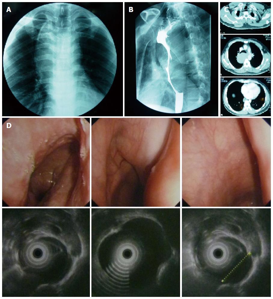
Figure 1 Mass showed before the surgery.
A: The frontal chest radiograph showed a widened and undulated mediastinal contour; B: Barium esophagogram demonstrated a multilobulated protuberant lesion with a relatively smooth margin in the middle thoracic esophagus; C: Chest CT scan revealed a 13 cm × 5 cm × 6 cm hyperdense soft-tissue mass, originated from the posterior wall of the esophagus. The mass was well demarcated from the surrounding structures protruded into the esophageal lumen and compressed the trachea and the right main bronchus, and no metastases noted was found; D: ESU demonstrates a hypoechoic, homogeneous, well-demarcated mass, arising at 24 cm from the incisors and extending to 35 cm and arising from the muscularis propria of the esophagus.
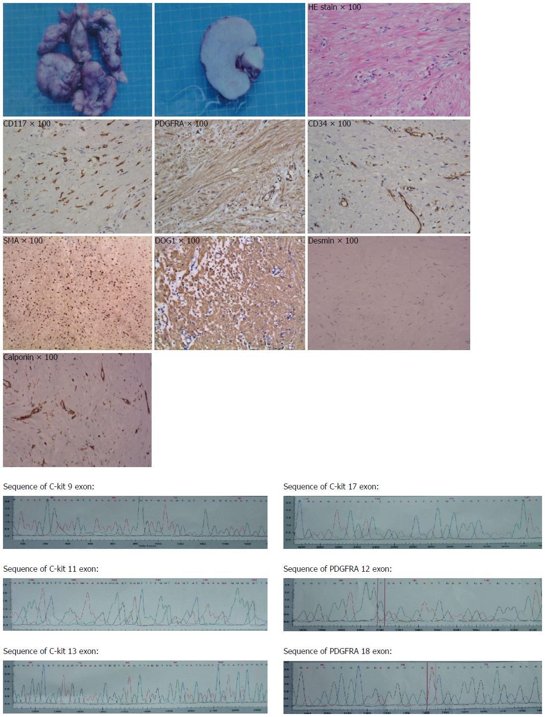
Figure 2 Pathology revealed a 13.
0 cm × 12.0 cm × 5.0 cm mass with an intact pseudocapsule. The tumor was CD117 (C-kit), PDGFRA, DOG1 and SMA positive, CD34 and calponinand weak positive, and negative for desmin. Mutation sequencing found that exon 12 and exon18 of PDGFRA and exon 9,11,13,17 of C-kit all were wild type.
- Citation: Mu ZM, Xie YC, Peng XX, Zhang H, Hui G, Wu H, Liu JX, Chen BK, Wu D, Ye YW. Long-term survival after enucleation of a giant esophageal gastrointestinal stromal tumor. World J Gastroenterol 2014; 20(37): 13632-13636
- URL: https://www.wjgnet.com/1007-9327/full/v20/i37/13632.htm
- DOI: https://dx.doi.org/10.3748/wjg.v20.i37.13632


