©2014 Baishideng Publishing Group Inc.
World J Gastroenterol. Oct 7, 2014; 20(37): 13591-13598
Published online Oct 7, 2014. doi: 10.3748/wjg.v20.i37.13591
Published online Oct 7, 2014. doi: 10.3748/wjg.v20.i37.13591
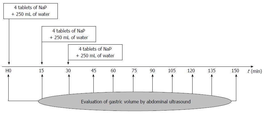
Figure 1 Protocol schedule the day of the colonoscopy.
NaP: Sodium phosphate bowel preparation.
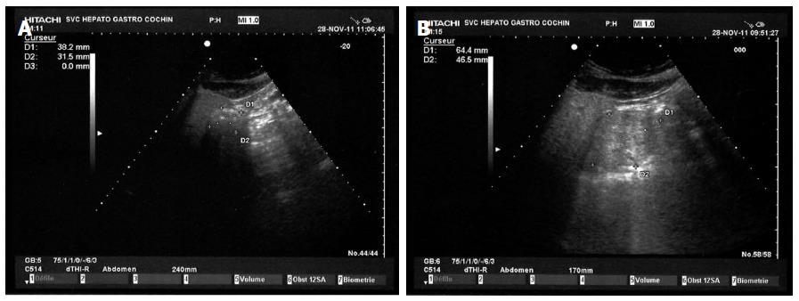
Figure 2 Measurement of the surface antral by ultrasonography in a patient in right lateral position at baseline.
A: Measurement after 12 h of gastric rest; B: Measurement after the ingestion of 750 mL water plus 12 tablets of NaP in 45 min showing a distortion of the antrum. NaP: Sodium phosphate bowel preparation.
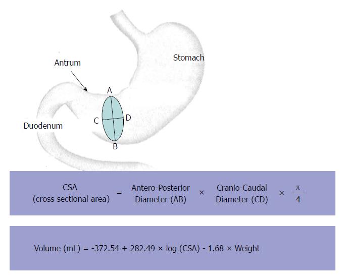
Figure 3 Ultrasound method of assessing gastric emptying time based on measurements of gastric antrum.
CSA: Cross sectional area.
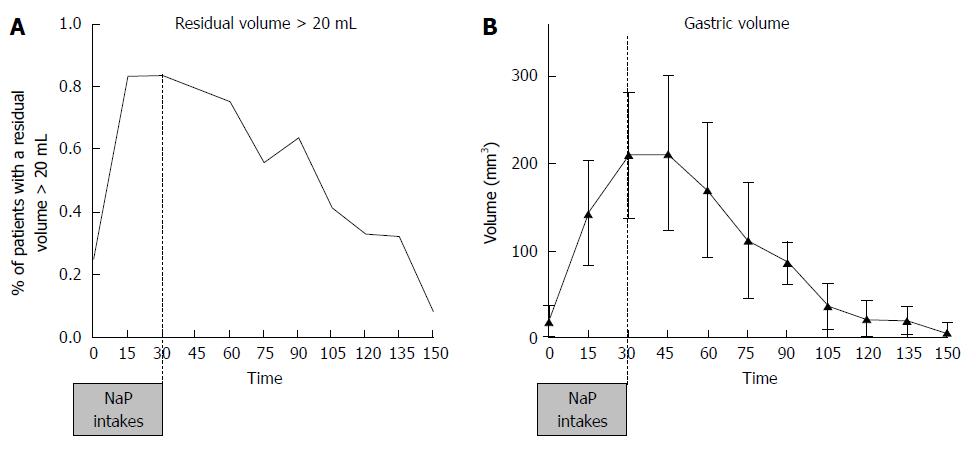
Figure 4 Evaluation of gastric volume and residual volume using ultrasonography.
A: Percent of patients with a residual antral volume above 20 mL at each time. Results are expressed in percent of the overall population (n = 27); B: Modification of the gastric volume. Results are expressed in mean ± SD of the overall population (n = 27).
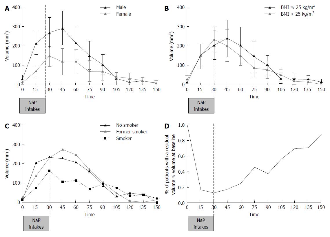
Figure 5 Evolution of gastric volume.
A: Evolution of gastric volume according to gender; B: Evolution of gastric volume according to body mass index; C: Evolution of gastric volume according to or smoking habits; D: Gastric emptying evolution considering the time when gastric volume returns to baseline (after 12 h fasting).
- Citation: Coriat R, Polin V, Oudjit A, Henri F, Dhooge M, Leblanc S, Delchambre C, Esch A, Tabouret T, Barret M, Prat F, Chaussade S. Gastric emptying evaluation by ultrasound prior colonoscopy: An easy tool following bowel preparation. World J Gastroenterol 2014; 20(37): 13591-13598
- URL: https://www.wjgnet.com/1007-9327/full/v20/i37/13591.htm
- DOI: https://dx.doi.org/10.3748/wjg.v20.i37.13591


