©2014 Baishideng Publishing Group Inc.
World J Gastroenterol. Sep 21, 2014; 20(35): 12704-12708
Published online Sep 21, 2014. doi: 10.3748/wjg.v20.i35.12704
Published online Sep 21, 2014. doi: 10.3748/wjg.v20.i35.12704
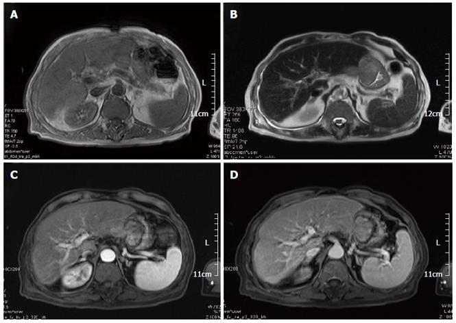
Figure 1 Magnetic resonance imaging.
A: T1-weighted imaging showing equisignal tumors in the left lateral hepatic lobe and Gastric lumen; B: T2-weighted imaging showing the tumor in the left lateral hepatic lobe; C, D: MR images showing slightly increased signals in the left lateral hepatic lobe and Gastric lumen.
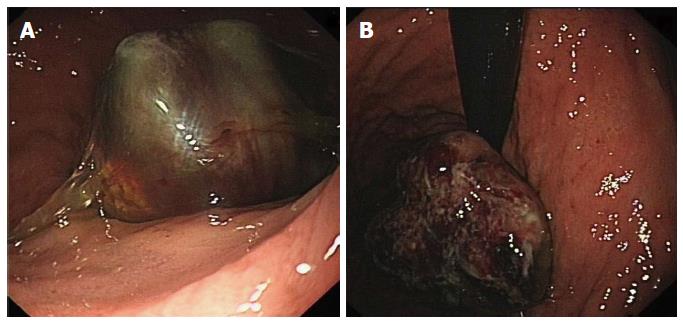
Figure 2 Gastroscopy imaging.
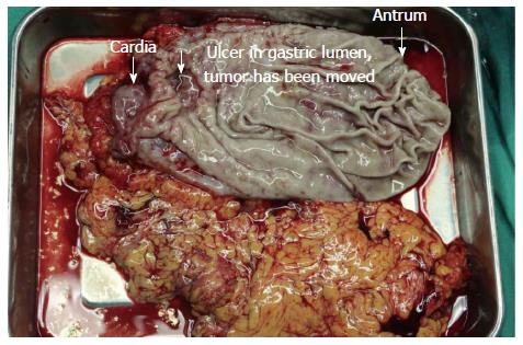
Figure 3 Intraoperative specimen.
Following removal of the gastric lumen tumor, an ulcer could be seen in the lesser curvature, 4 cm away from the cardia.
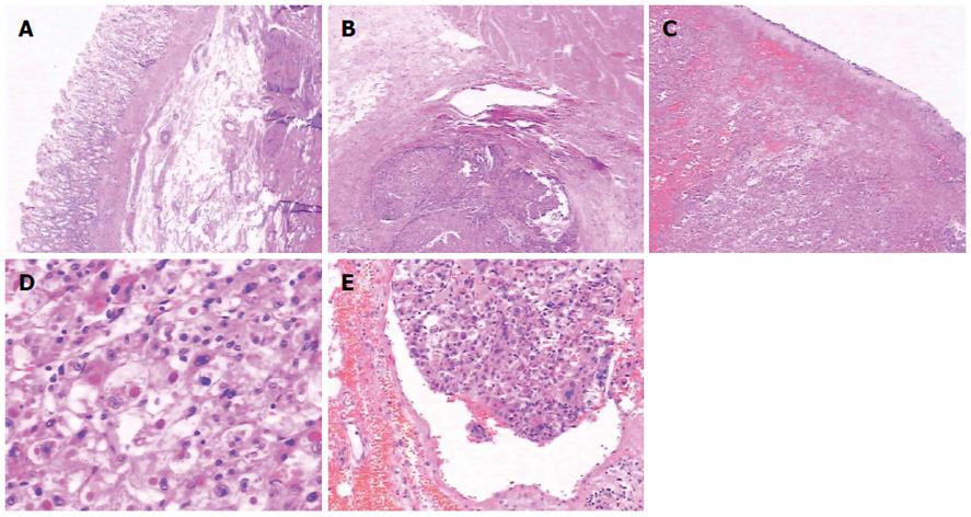
Figure 4 Pathological results (HE staining).
A: × 40, mucosa was intact; B: × 40, tumor was mainly localized in saroas; C: × 20, tumor cells in gastric lumen was necrosis with bleeding; D: × 200, tumor cells distributed like hepatic sinusoid; E: × 100, tumor embolus could be seen in vessels.
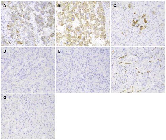
Figure 5 Immunohistochemical results.
A: CK8/18; B: Hepar-1+; C: CK 20 focal+; D: AFP-; E: CEA-; F: CD34 vessel; G: CD117-.
- Citation: Wu WD, Wu J, Yang HG, Chen Y, Zhang CW, Zhao DJ, Hu ZM. Rare cause of upper gastrointestinal bleeding owing to hepatic cancer invasion: A case report. World J Gastroenterol 2014; 20(35): 12704-12708
- URL: https://www.wjgnet.com/1007-9327/full/v20/i35/12704.htm
- DOI: https://dx.doi.org/10.3748/wjg.v20.i35.12704


