Copyright
©2014 Baishideng Publishing Group Inc.
World J Gastroenterol. Jul 28, 2014; 20(28): 9626-9630
Published online Jul 28, 2014. doi: 10.3748/wjg.v20.i28.9626
Published online Jul 28, 2014. doi: 10.3748/wjg.v20.i28.9626
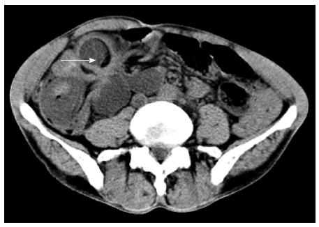
Figure 1 Computed tomography scan showing right lower intestinal intussusception (arrow) and multiple enlarged lymph nodes within the abdominal cavity and retroperitoneum.
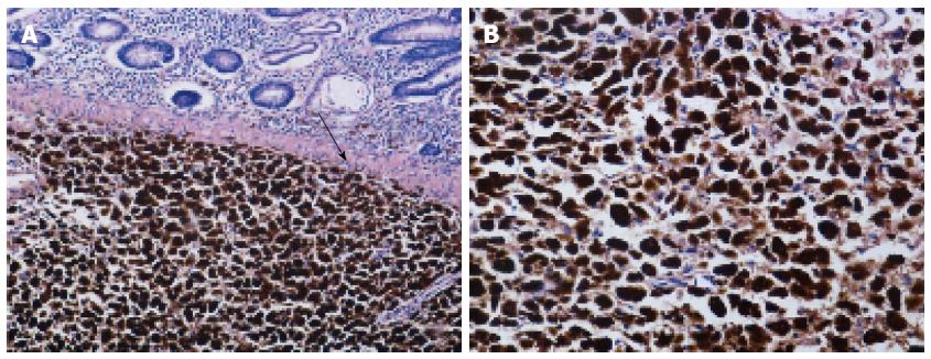
Figure 2 Histopathologic findings of excised specimen.
A: Microscopic examination showing tumor cells invading the colonic submucosa (arrow) (hematoxylin and eosin, × 100); B: Microscopic examination showing epithelioid and spindle tumor cells with melanin deposition in the cytoplasm (hematoxylin and eosin, × 200).
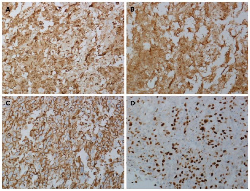
Figure 3 Immunohistochemistry findings of excised specimen.
A-C: Tumor cells diffusely expressing S-100 (A), HMB-45 (B) and vimentin (C); D: 80% of tumor cells were positive for Ki-67 (immunohistochemistry, × 200).
- Citation: Li WX, Wei Y, Jiang Y, Liu YL, Ren L, Zhong YS, Ye LC, Zhu DX, Niu WX, Qin XY, Xu JM. Primary colonic melanoma presenting as ileocecal intussusception: Case report and literature review. World J Gastroenterol 2014; 20(28): 9626-9630
- URL: https://www.wjgnet.com/1007-9327/full/v20/i28/9626.htm
- DOI: https://dx.doi.org/10.3748/wjg.v20.i28.9626


