©2014 Baishideng Publishing Group Inc.
World J Gastroenterol. Jul 14, 2014; 20(26): 8736-8739
Published online Jul 14, 2014. doi: 10.3748/wjg.v20.i26.8736
Published online Jul 14, 2014. doi: 10.3748/wjg.v20.i26.8736
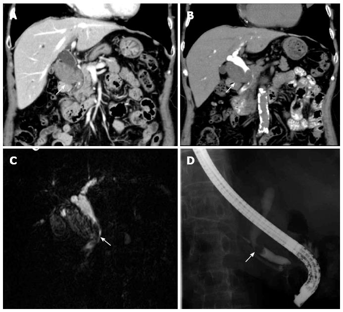
Figure 1 A 79-year-old female presented to our hospital for an incidentally-diagnosed gallbladder tumor.
A: Coronal contrast-enhanced computed tomography (CT) image. The white arrow points to the gallbladder tumor with bile duct tumor thrombus; B: Coronal drip infusion cholangiographic CT image. The white arrow indicates the gallbladder tumor with bile duct tumor thrombus; C: Endoscopic retrograde cholangiopancreatography image. The white arrow indicates the defect due to the bile duct tumor thrombus; D: Magnetic resonance cholangiopancreatography image. The white arrow indicates the defect due to the bile duct tumor thrombus.
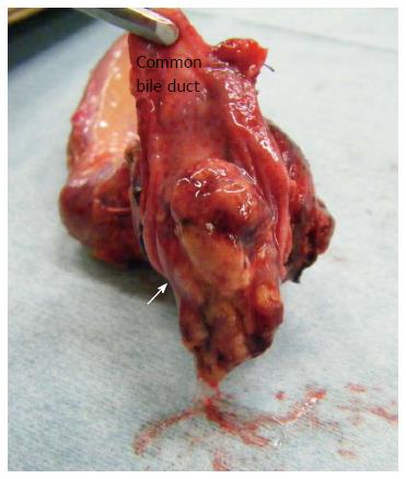
Figure 2 The resected specimen.
Dissection of the common bile duct demonstrated a mucin-producing tumor thrombus.
Figure 3 Pathological examination of the tumor.
A: Hematoxylin and eosin staining demonstrated the tumor to be a tubulopapillary adenoma with moderate epithelial atypia (× 400); B: MUC5AC staining showing positive expression (× 600).
- Citation: Yamamoto K, Yamamoto F, Maeda A, Igimi H, Yamamoto M, Yamaguchi R, Yamashita Y. Tubulopapillary adenoma of the gallbladder accompanied by bile duct tumor thrombus. World J Gastroenterol 2014; 20(26): 8736-8739
- URL: https://www.wjgnet.com/1007-9327/full/v20/i26/8736.htm
- DOI: https://dx.doi.org/10.3748/wjg.v20.i26.8736


