Copyright
©2014 Baishideng Publishing Group Co.
World J Gastroenterol. Apr 28, 2014; 20(16): 4718-4722
Published online Apr 28, 2014. doi: 10.3748/wjg.v20.i16.4718
Published online Apr 28, 2014. doi: 10.3748/wjg.v20.i16.4718
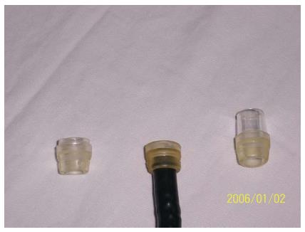
Figure 1 Improved transparent cap.
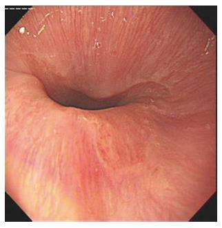
Figure 2 Tongue-type Barrett’s esophagus (without transparent cap).
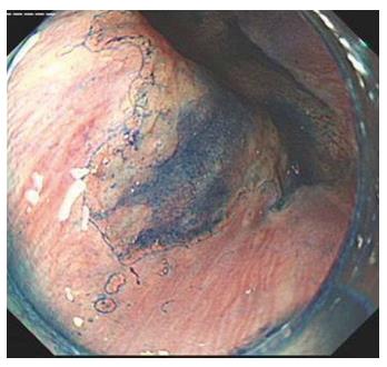
Figure 3 Same lesion as in Figure 1 (with transparent cap).
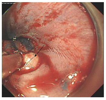
Figure 4 Targeted biopsy of the suspected Barrett’s esophagus lesion (the same lesion as in Figure 1).
The Z line is indicated by the arrow.
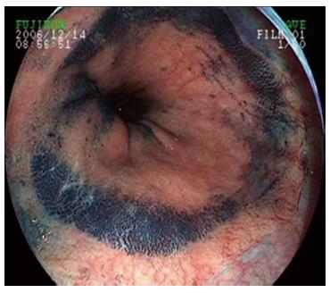
Figure 5 Intestinal metaplasia in circumferential-type Barrett’s esophagus, indicated by methylene blue dye.
- Citation: Chen BL, Xing XB, Wang JH, Feng T, Xiong LS, Wang JP, Cui Y. Improved biopsy accuracy in Barrett’s esophagus with a transparent cap. World J Gastroenterol 2014; 20(16): 4718-4722
- URL: https://www.wjgnet.com/1007-9327/full/v20/i16/4718.htm
- DOI: https://dx.doi.org/10.3748/wjg.v20.i16.4718


