©2014 Baishideng Publishing Group Co.
World J Gastroenterol. Apr 14, 2014; 20(14): 4030-4036
Published online Apr 14, 2014. doi: 10.3748/wjg.v20.i14.4030
Published online Apr 14, 2014. doi: 10.3748/wjg.v20.i14.4030
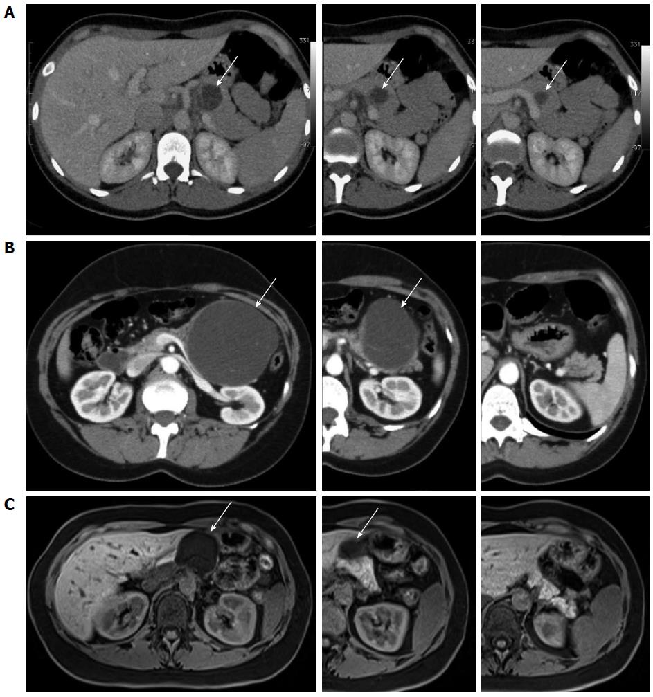
Figure 1 Abdominal computed tomography scan.
A: Abdominal computed tomography (CT) scan showing a 4 cm pancreatic cystic lesion of the neck in patient 1; B: Abdominal CT scan showing a 10 cm pancreatic cystic lesion of the body in patient 2; C: Abdominal magnetic resonance imaging showing a 4.6 cm cystic lesion of the neck in patient 3. Arrows mark the cystic lesion (A-C).
Figure 2 Specimen after distal pancreatic resection.
A: An arrow marks the cystic lesion; the interrupted line marks the site of transection; B: Transection of the pancreas distal from the lesion; C: Spared pancreatic parenchyma.
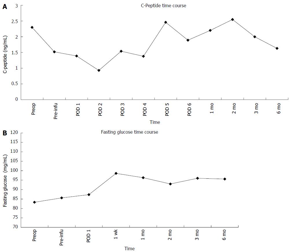
Figure 3 Fasting C-peptide (A) and fasting glycemia (B) remained stable and comparable with respect to pretransplant value, respectively.
The values are expressed as mean plus the standard error of the mean. Preop: Preoperative value; Preinfu: C-peptide value after resection and before islet re-infusion; POD: Post operative day.
- Citation: Balzano G, Carvello M, Piemonti L, Nano R, Ariotti R, Mercalli A, Melzi R, Maffi P, Braga M, Staudacher C. Combined laparoscopic spleen-preserving distal pancreatectomy and islet autotransplantation for benign pancreatic neoplasm. World J Gastroenterol 2014; 20(14): 4030-4036
- URL: https://www.wjgnet.com/1007-9327/full/v20/i14/4030.htm
- DOI: https://dx.doi.org/10.3748/wjg.v20.i14.4030


