©The Author(s) 1996.
World J Gastroenterol. Sep 15, 1996; 2(3): 152-154
Published online Sep 15, 1996. doi: 10.3748/wjg.v2.i3.152
Published online Sep 15, 1996. doi: 10.3748/wjg.v2.i3.152
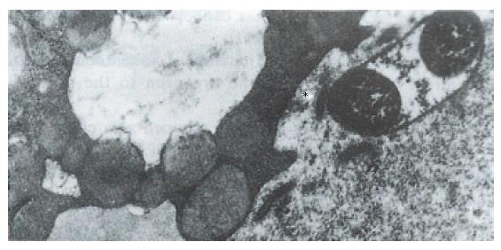
Figure 1 The bacillus abuts upon the depression of the plasma membrane (*) of the vacuolated cell.
( × 21000)
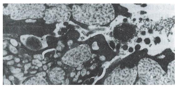
Figure 2 The bacilli (*) penetrating deep in between the mucous cells.
The tight junction was broken and the intercellular space was dilated. ( × 21000)
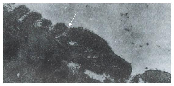
Figure 3 The bacillus shows the first step of adherence initiated by the direct contact of the organism to the microvilla glycocalyx.
( × 28500)
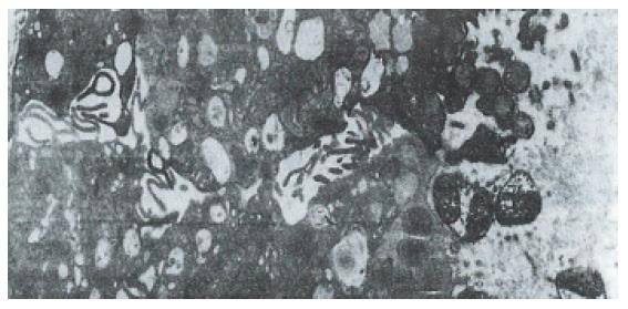
Figure 4 A great number of bacteria grouped in a colony shown with periplasmic pools (s).
The bacilli abutting upon the depression of the plasma membrane of mucous neck cells lost microvilli and vacuolated. ( × 8700)
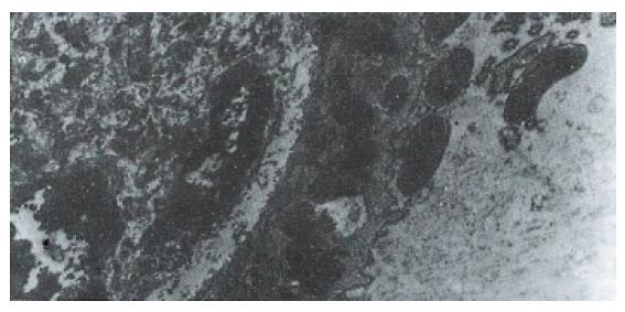
Figure 5 Neutrophils (*) penetrated into the epithelia and migrated to the bacilli located in the apical region of mucous neck cells.
( × 10800)
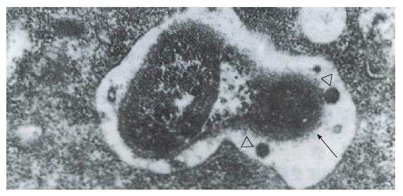
Figure 6 The bacillus is phagocytized by neutrophils located in the lumen.
In the phagosome, two lysosomes approach the bacillus, of which the cell wall was lysed and discontinuous. ( × 43500)
- Citation: Yang SM, Lin BZ, Fang Y, Zheng Y. Ultrastructural observation of Helicobacter pylori to the gastric epithelia in chronic gastritis and peptic ulcers. World J Gastroenterol 1996; 2(3): 152-154
- URL: https://www.wjgnet.com/1007-9327/full/v2/i3/152.htm
- DOI: https://dx.doi.org/10.3748/wjg.v2.i3.152


