©2013 Baishideng Publishing Group Co.
World J Gastroenterol. Mar 7, 2013; 19(9): 1485-1488
Published online Mar 7, 2013. doi: 10.3748/wjg.v19.i9.1485
Published online Mar 7, 2013. doi: 10.3748/wjg.v19.i9.1485
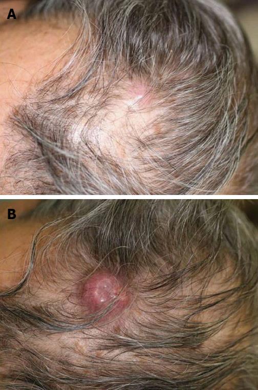
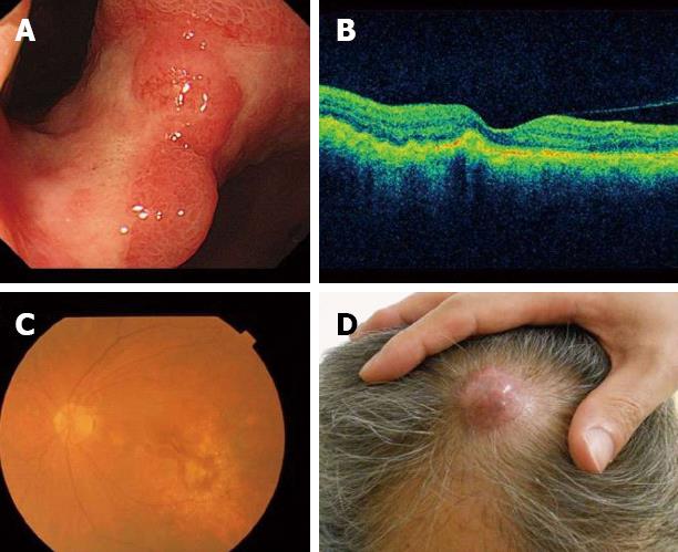
Figure 1 Cutaneous metastasis lesion in the head.
A: One of the two cutaneous nodules on his head at first visit; B: The size of the cutaneous tumor was rapidly enlarged at 1 mo after the first visit.
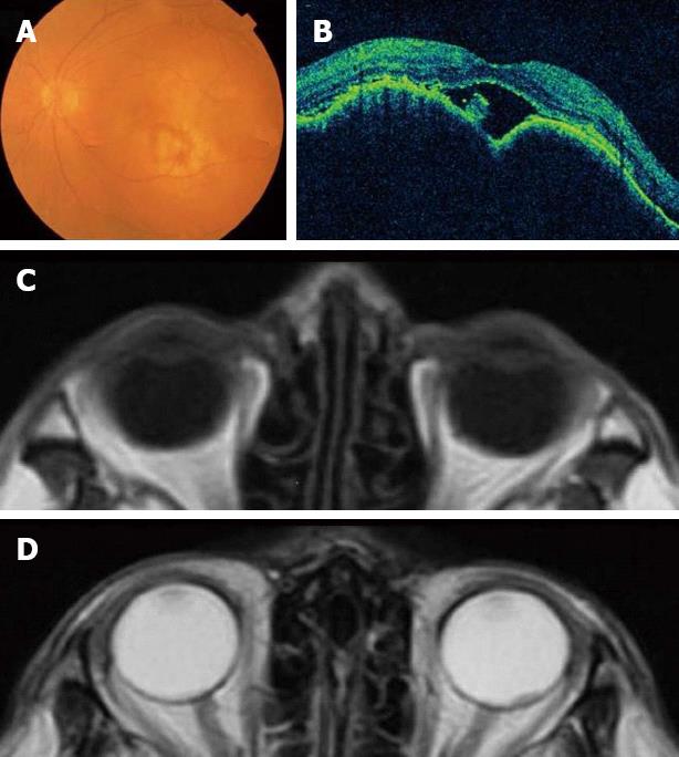
Figure 2 An elevated choroidal neoplasm.
A: Fundoscopic examination; B: Optical coherence tomographic examination; C: Magnetic resonance imaging (MRI) T1-weighted image; D: MRI T2-weighted image.
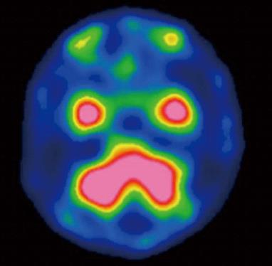
Figure 3 N-isopropyl-p-(123I) iodoamphetamine single photon emission computed tomography findings.
123 Iodoamphetamine single photon emission computed tomography showed no accumulation in bilateral eyes.
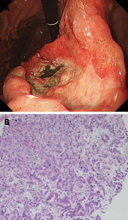
Figure 4 Esophagogastroduodenoscopy and pathological findings of gastric cancer.
A: Esophagogastroduodenoscopy was performed and revealed an advanced gastric cancer; B: The gastric biopsy specimens revealed moderately differentiated adenocarcinoma (hematoxylin and eosin stain, ×400).
Figure 5 Response to chemotherapy.
A: The primary gastric lesion was markedly reduced after 2 cycles of chemotherapy; B, C: The choroidal metastatic tumor was also markedly reduced; D: The larger cutaneous nodule of head enlarged from 25 mm to 32 mm, and headache vanished.
- Citation: Kawai S, Nishida T, Hayashi Y, Ezaki H, Yamada T, Shinzaki S, Miyazaki M, Nakai K, Yakushijin T, Watabe K, Iijima H, Tsujii M, Nishida K, Takehara T. Choroidal and cutaneous metastasis from gastric adenocarcinoma. World J Gastroenterol 2013; 19(9): 1485-1488
- URL: https://www.wjgnet.com/1007-9327/full/v19/i9/1485.htm
- DOI: https://dx.doi.org/10.3748/wjg.v19.i9.1485


