Copyright
©2013 Baishideng Publishing Group Co.
World J Gastroenterol. Mar 7, 2013; 19(9): 1458-1465
Published online Mar 7, 2013. doi: 10.3748/wjg.v19.i9.1458
Published online Mar 7, 2013. doi: 10.3748/wjg.v19.i9.1458
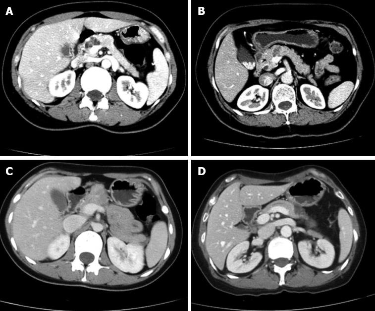
Figure 1 Location of lesions in the pancreas.
A: Head-neck; B: Neck; C: Neck-body; D: Body.
Figure 2 The surgical approach of the cephalic pancreatic cut surface.
A: Closed by Endo-GIA™ 60-2.5 auto suture; B: Continuous suture using 4-0 prolene;
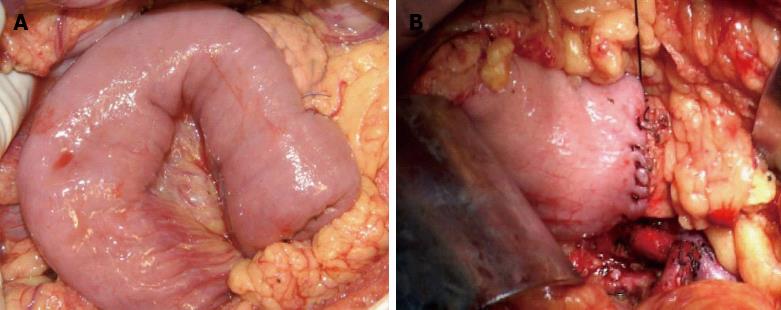
Figure 3 The reconstruction of the distal side stump.
A: Pancreaticojejunostomy; B: Pancreaticogastrostomy.
- Citation: Du ZY, Chen S, Han BS, Shen BY, Liu YB, Peng CH. Middle segmental pancreatectomy: A safe and organ-preserving option for benign and low-grade malignant lesions. World J Gastroenterol 2013; 19(9): 1458-1465
- URL: https://www.wjgnet.com/1007-9327/full/v19/i9/1458.htm
- DOI: https://dx.doi.org/10.3748/wjg.v19.i9.1458


