©2013 Baishideng Publishing Group Co.
World J Gastroenterol. Dec 28, 2013; 19(48): 9481-9484
Published online Dec 28, 2013. doi: 10.3748/wjg.v19.i48.9481
Published online Dec 28, 2013. doi: 10.3748/wjg.v19.i48.9481
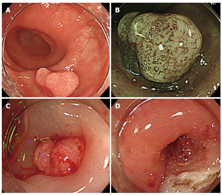
Figure 1 Endoscopic mucosal dissection of the sigmoid colon polyp.
A: A sigmoid colon polyp approximately 25 mm in diameter; B: Narrow band imaging magnified colonoscopy was performed to investigate the polyp in greater detail. Several irregular microvessels were observed on the surface of the polyp, but there was no pit pattern on the surface; C: A local saline injection was administered, and we observed slight elevation of the polyp; D: After the endoscopic mucosal resection procedure and the removal of the polyp, the diverticulum was identified using the resected stalk of the polyp.
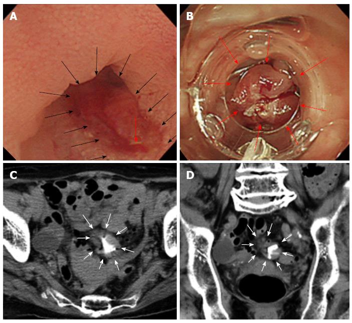
Figure 2 Closure of the diverticulum using the resected stalk of the polyp.
A: In a closer view of the resected surface, the cavity of the diverticulum was irregular (the black arrows), and an exposed vessel was identified (red arrow); resection of the polyp indicated that it arose from the diverticulum; B: To prevent bleeding and delayed perforation following the endoscopic mucosal resection (EMR) procedure, we inverted the diverticulum and sutured the inverted diverticulum, including the resected stalk of the polyp, using an over-the-scope clip (red arrows); C, D: After the EMR procedure, computed tomography was performed to examine the soft tissue density around the over-the-scope clip and the increased fat density around the resected site (white arrows).
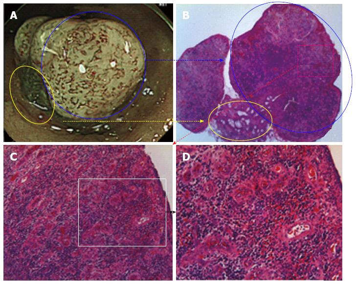
Figure 3 Histological findings of the resected polyp.
A: A 20 magnified narrow band imaging image of the granulomatous polyp; B: A 20 magnified image with a hematoxylin and eosin (HE) stain, the yellow and blue circles in Panel A corresponding to those in Panel B; C: A 100 magnified image with a HE stain reveals significant infiltration of lymphocytes and plasma cells; D: A 200 magnified image with a HE stain reveals increased outgrowth of microvascular structures and infiltration of lymphocytes, neutrophils and plasma cells, which indicates granulation tissue. There were no atypical cells or structural atypia.
- Citation: Mori H, Tsushimi T, Kobara H, Nishiyama N, Fujihara S, Matsunaga T, Ayagi M, Yachida T, Masaki T. Endoscopic management of a rare granulation polyp in a colonic diverticulum. World J Gastroenterol 2013; 19(48): 9481-9484
- URL: https://www.wjgnet.com/1007-9327/full/v19/i48/9481.htm
- DOI: https://dx.doi.org/10.3748/wjg.v19.i48.9481


