Copyright
©2013 Baishideng Publishing Group Co.
World J Gastroenterol. Dec 28, 2013; 19(48): 9392-9398
Published online Dec 28, 2013. doi: 10.3748/wjg.v19.i48.9392
Published online Dec 28, 2013. doi: 10.3748/wjg.v19.i48.9392
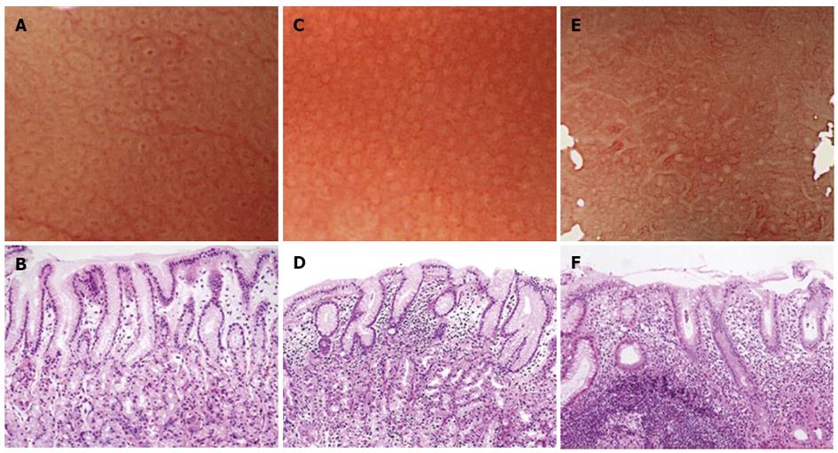
Figure 1 The whiteness of crypt openings (HE, × 100).
A: "White-edged dark spot" crypt openings (COs) (a round dark spot bordered by white); B: Histological image of "white-edged dark spot" COs; C: "White" COs (pure white COs without a dark spot); D: Histological image of "white" COs; E: "Dense white pit" ("DWP") COs (densely white COs resembling snowballs); F: Histological image of "DWP" COs.
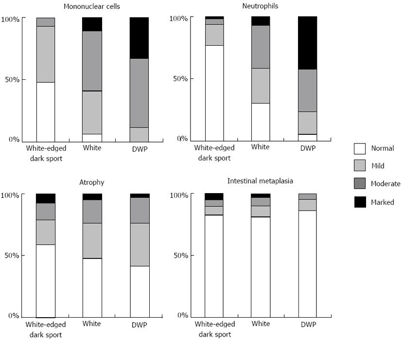
Figure 2 Whiteness of crypt opening and grade of gastritis according to the updated Sydney System.
Kruskal-Wallis test showed significant correlations between grades of histological inflammation (P < 0.001) and activity (P < 0.001) and whiteness of crypt openings (COs). Glandular atrophy and intestinal metaplasia were not significantly correlated with whiteness of COs. DWP: Dense white pit.
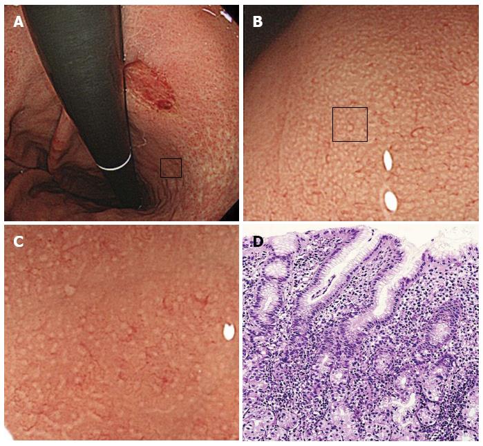
Figure 3 A case with an early undifferentiated-type gastric cancer in the middle of the corpus.
A: A 67-year-old woman was found to have a depressed undifferentiated-type gastric cancer about 20 mm in diameter in an area found endoscopically to be non-atrophic; B: Magnifying endoscopy image showing minute round pits in uninvolved corpus mucosa; C: The whiteness of the crypt openings is "dense white pit" type; they resemble snowballs; D: Microscopic examination of uninvolved corpus mucosa revealed marked lymphocyte and neutrophil infiltration in the lamina propria and neutrophil infiltration within the foveolar lumens (HE stain, × 100).
-
Citation: Kawamura M, Sekine H, Abe S, Shibuya D, Kato K, Masuda T. Clinical significance of white gastric crypt openings observed
via magnifying endoscopy. World J Gastroenterol 2013; 19(48): 9392-9398 - URL: https://www.wjgnet.com/1007-9327/full/v19/i48/9392.htm
- DOI: https://dx.doi.org/10.3748/wjg.v19.i48.9392


