©2013 Baishideng Publishing Group Co.
World J Gastroenterol. Nov 28, 2013; 19(44): 8065-8070
Published online Nov 28, 2013. doi: 10.3748/wjg.v19.i44.8065
Published online Nov 28, 2013. doi: 10.3748/wjg.v19.i44.8065
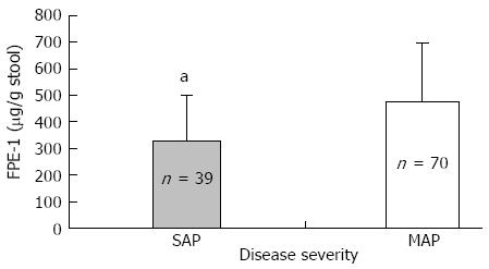
Figure 1 Disease severity and fecal pancreatic elastase-I level.
Fecal pancreatic elastase-I (FPE-1) level was lower in severe acute pancreatitis (SAP) than mild acute pancreatitis (MAP) (aP < 0.05). FPE levels are 330.9 ± 170.6 μg/g and 475.2 ± 223 μg/g stool in SAP and MAP, respectively.
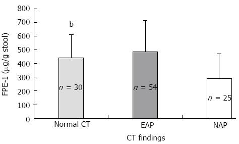
Figure 2 Fecal pancreatic elastase-I levels and contrast enhanced abdominal computed tomography findings.
Fecal pancreatic elastase-I (FPE-1) level was significantly lower in necrotizing acute pancreatitis (NAP) than the patients without pancreatic necrosis (bP < 0.001). FPE-I levels as follows; 292.43 ± 178.78, 487 ± 226.21, and 443.91 ± 167.83 μg/g stool in NAP, edematous acute pancreatitis (EAP) and normal contrast enhanced abdominal computed tomography (CT) groups, respectively.
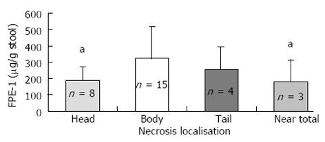
Figure 3 Necrosis localization and fecal pancreatic elastase-I levels.
Fecal pancreatic elastase-I (FPE-1) level was lower in head and near total necrosis of the pancreas than body or tail necrosis (aP < 0.05). FPE–1 levels as follows; 189.18 ± 82.7, 324.86 ± 194, 258.06 ± 134.63 and 181.12 ± 134.25 μg/g stool in head, body, tail and near total necrosis groups, respectively.
Figure 4 Acute pancreatitis and endocrine dysfunction.
Endocrine dysfunction was much higher in severe acute pancreatitis than mild acute pancreatitis (A) and also much higher in necrotizing acute pancreatitis than without necrosis (B) (bP < 0.001). SAP: Severe acute pancreatitis; MAP: Mild acute pancreatitis; NAP: Necrotizing acute Pancreatitis; EAP: Edematous acute pancreatitis.
- Citation: Garip G, Sarandöl E, Kaya E. Effects of disease severity and necrosis on pancreatic dysfunction after acute pancreatitis. World J Gastroenterol 2013; 19(44): 8065-8070
- URL: https://www.wjgnet.com/1007-9327/full/v19/i44/8065.htm
- DOI: https://dx.doi.org/10.3748/wjg.v19.i44.8065


