©2013 Baishideng Publishing Group Co.
World J Gastroenterol. Sep 21, 2013; 19(35): 5837-5847
Published online Sep 21, 2013. doi: 10.3748/wjg.v19.i35.5837
Published online Sep 21, 2013. doi: 10.3748/wjg.v19.i35.5837
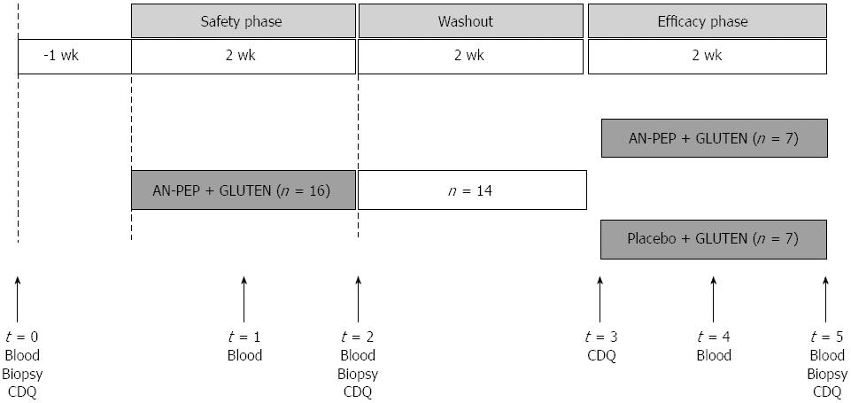
Figure 1 Study design and flowchart.
In the safety phase, 16 patients daily consumed 5 pieces of toast with Aspergillus niger prolyl endoprotease (AN-PEP) for 2 wk while continuing their gluten-free diet (GFD). Two patients deteriorated on Marsh scores and were excluded. After a 2-wk wash-out period during which the patients continued their usual GFD, the remainder of 14 patients were randomized to the efficacy phase to receive 2 wk of toast with either AN-PEP or placebo while continuing their GFD. CDQ: Celiac disease quality.
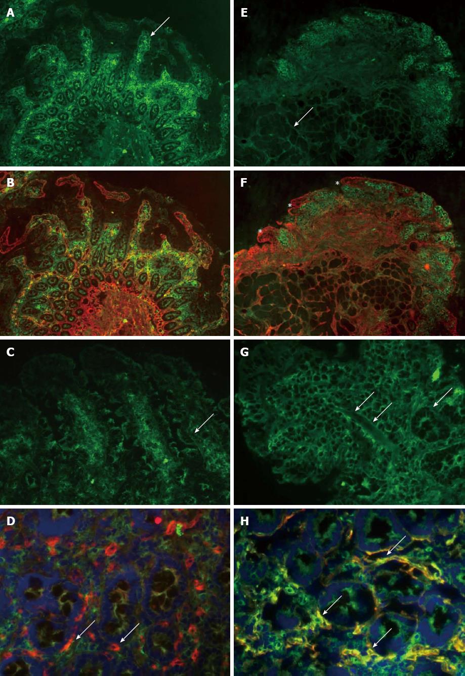
Figure 2 Small intestinal tissue transglutaminase IgA antibody deposits (rated on a scale 0-3) in two patients at baseline and after randomization to Aspergillus niger prolyl endoprotease and placebo respectively.
A: Baseline evaluation of patient 1 showed preserved villous architecture (arrow), with intense, grade 3 IgA depositions (green) subepithelially and around crypts; B: This deposition merges to yellow indicating co-localisation with tTG shown in red; C: In this patient, IgA deposits diminished after 2 wk Aspergillus niger prolyl endoprotease (AN-PEP) treatment to grade 1, when only faint and patchy antibody deposition was seen (arrow); D: tTG appeared in red in this AN-PEP-treated patient (arrow) in the absence of IgA deposition. The cell nuclei were stained with 4',6-diamidino-2-phenylindole (DAPI) (blue); E: Baseline evaluation of patient 2 showed preserved villous architecture with faint, grade 1 IgA deposition in the deep mucosal layer around Brunner glands (arrow); F: This deposition were not sufficient to obtain a yellow colour at merging with tTG shown in red (asterisk); G: In this patient, IgA deposition increased to grade 2 subepithelially (arrow) after 2 wk placebo; H: IgA deposition increased to grade 3 in the crypt region after 2-wk placebo (arrow). IgA deposition co-localised with tTG to intense yellow (arrow). The cell nuclei were stained with DAPI (blue).
- Citation: Tack GJ, van de Water JM, Bruins MJ, Kooy-Winkelaar EM, van Bergen J, Bonnet P, Vreugdenhil AC, Korponay-Szabo I, Edens L, von Blomberg BME, Schreurs MW, Mulder CJ, Koning F. Consumption of gluten with gluten-degrading enzyme by celiac patients: A pilot-study. World J Gastroenterol 2013; 19(35): 5837-5847
- URL: https://www.wjgnet.com/1007-9327/full/v19/i35/5837.htm
- DOI: https://dx.doi.org/10.3748/wjg.v19.i35.5837


