Copyright
©2013 Baishideng Publishing Group Co.
World J Gastroenterol. Aug 14, 2013; 19(30): 5025-5028
Published online Aug 14, 2013. doi: 10.3748/wjg.v19.i30.5025
Published online Aug 14, 2013. doi: 10.3748/wjg.v19.i30.5025
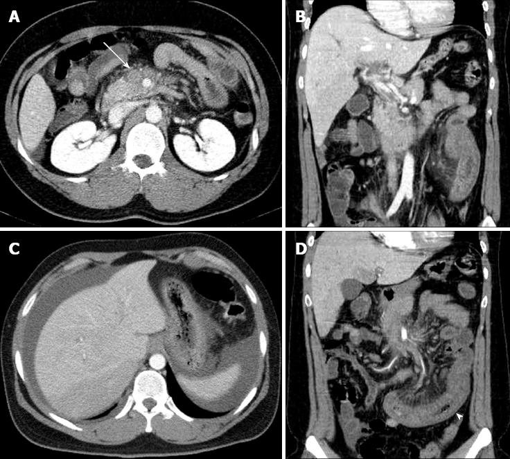
Figure 1 Abdominal computed tomography demonstrates an acute mesenteric venous thrombosis at the time of initial presentation.
A: A thrombus (arrow) and perivenous infiltration at the proximal superior mesenteric vein; B: extension into the portal vein; C: An abnormal fluid collection around the liver and spleen; D: The affected small bowel (arrow head) with long-segment wall thickening and decreased enhancement.
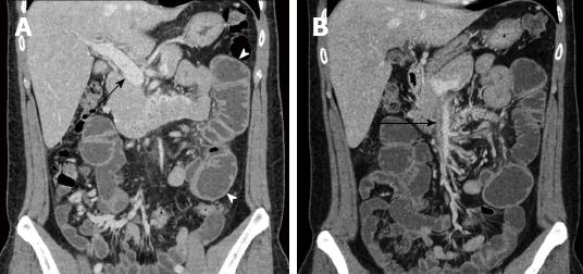
Figure 2 Abdominal computed tomography at second admission.
A: Dilated proximal jejunal loop (arrow heads) and resolution of thrombus in the main portal vein (arrow); B: Remnant thrombus in superior mesenteric vein (arrow).
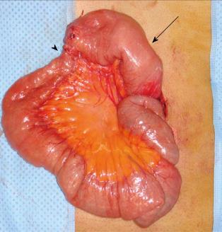
Figure 3 Intraoperative findings: sequelae of the mesenteric venous thrombosis.
A dilated proximal jejunum (arrow) and short-segment stricture (arrow head) are noted.
- Citation: Kim HK, Chun JM, Huh S. Anticoagulation and delayed bowel resection in the management of mesenteric venous thrombosis. World J Gastroenterol 2013; 19(30): 5025-5028
- URL: https://www.wjgnet.com/1007-9327/full/v19/i30/5025.htm
- DOI: https://dx.doi.org/10.3748/wjg.v19.i30.5025


