Copyright
©2013 Baishideng Publishing Group Co.
World J Gastroenterol. Aug 14, 2013; 19(30): 4925-4934
Published online Aug 14, 2013. doi: 10.3748/wjg.v19.i30.4925
Published online Aug 14, 2013. doi: 10.3748/wjg.v19.i30.4925
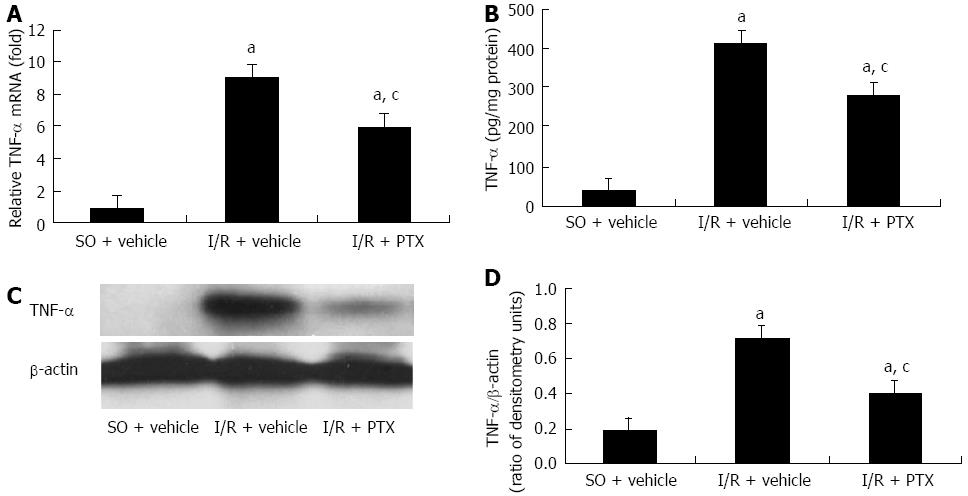
Figure 1 Ischemia-reperfusion induced intestinal mucosal tumor necrosis factor-α expression.
A: Small intestinal mucosal tumor necrosis factor-α (TNF-α) mRNA expression; B: ELISA analysis of small intestinal mucosal TNF-α protein expression; C: Western blotting analysis of small intestinal mucosal TNF-α protein expression; D: The results of Western blotting analysis were expressed as a ratio to β-actin densitometry units. A TNF-α inhibitor, pentoxifylline (PTX), significantly suppressed the TNF-α mRNA and protein expression. Values are mean ± SE. Six rats were tested in each group. aP < 0.05 vs sham-operation (SO) rats pretreated with vehicle (SO + vehicle), cP < 0.05 vs ischemia-reperfusion (I/R) rats pretreated with vehicle (I/R + vehicle).
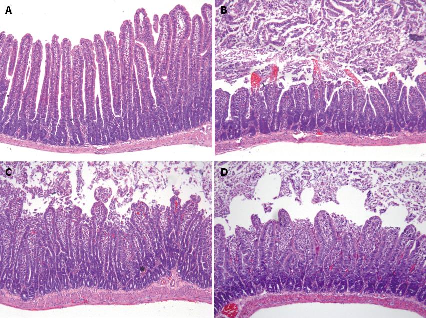
Figure 2 Suppression of tumor necrosis factor-α alleviated ischemia-reperfusion-induced small intestinal injury.
A: Sham-operation pretreated with vehicle; B: Ischemia-reperfusion (I/R) pretreated with vehicle. C: I/R pretreated with a tumor necrosis factor-α (TNF-α) inhibitor pentoxifylline; D: I/R pretreated with a TNF-α antibody infliximab; Representative sections of jejunum for hematoxylin and eosin staining (× 100) were showed. Six rats were studied in each group, and a similar pattern was seen in six different rats in each group.
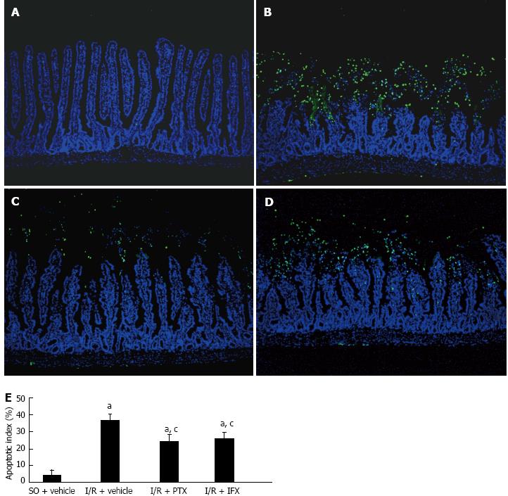
Figure 3 Suppression of tumor necrosis factor-α attenuated ischemia-reperfusion-induced intestinal mucosal apoptosis.
A: Sham-operation (SO) pretreated with vehicle; B: Ischemia-reperfusion (I/R) pretreated with vehicle; C: I/R pretreated with a tumor necrosis factor-α (TNF-α) inhibitor pentoxifylline (PTX); D: I/R pretreated with a TNF-α antibody infliximab (IFX); E: The apoptotic index was calculated by counting a minimum of 20 randomly selected villi and crypts in the sections following terminal deoxynucleotidyl transferase-mediated dUTP-biotin nick end labeling (TUNEL) staining. The index was obtained by dividing the TUNEL positive cells by the total number of cells. Values are mean ± SE. Six rats were tested in each group. aP < 0.05 vs SO rats pretreated with vehicle (SO + vehicle); cP < 0.05 vs I/R rats pretreated with vehicle (I/R + vehicle). Apoptosis was assessed by TUNEL immunofluorescence staining. Mid-jejunum sections of rats were stained by TUNEL (green), with nuclei counterstained by 4’,6-diamidino-2-phenylindole dihydrochloride (blue). Magnifications: × 100. Six rats were studied in each group, and a similar pattern was seen in six different rats in each group.
Figure 4 Suppression of tumor necrosis factor-α repressed caspase-3 activity in ischemia-reperfusion intestinal mucosa.
A: Sham-operation (SO) pretreated with vehicle; B: Ischemia-reperfusion (I/R) pretreated with vehicle; C: I/R pretreated with a tumor necrosis factor-α (TNF-α) inhibitor pentoxifylline (PTX); D: I/R pretreated with a TNF-α antibody infliximab (IFX); Activated caspase-3 was assessed by immunohistochemical staining. Magnifications: × 100; E: The active caspase-3 index was calculated by counting a minimum of 20 randomly selected villi and crypts in the sections following cleaved caspase-3 staining. The index was obtained by dividing the cleaved caspase-3 positive cells by the total number of cells. Values are mean ± SE. Six rats were tested in each group. aP < 0.05 vs SO rats pretreated with vehicle (SO + vehicle); cP < 0.05 vs I/R rats pretreated with vehicle (I/R + vehicle). Six rats were studied in each group, and a similar pattern was seen in six different rats in each group.
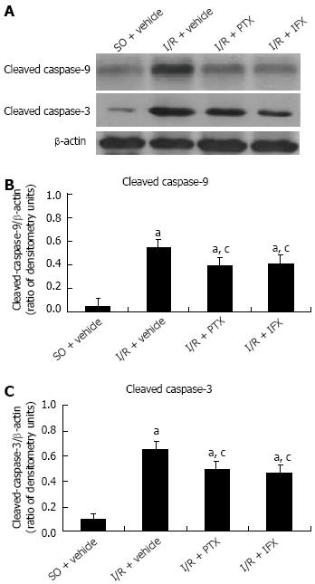
Figure 5 Tumor necrosis factor-α mediated ischemia-reperfusion-induced mucosal apoptosis via caspase activation in intestinal mucosa.
Total protein was extracted from intestinal mucosa, and subjected to SDS-PAGE and Western blotting analysis. β-actin was used as the control for loading. The results were expressed as a ratio to β-actin densitometry units. A: Western blotting analysis; B: The ratio of cleaved caspase-9 and β-actin; C: The ratio of cleaved caspase-3 and β-actin. Values are mean ± SE. Six rats were tested in each group. aP < 0.05 vs sham-operation (SO) rats pretreated with vehicle (SO + vehicle); cP < 0.05 vs ischemia-reperfusion(I/R) rats pretreated with vehicle (ischemia-reperfusion + vehicle). PTX: Pentoxifylline; IFX: Infliximab.
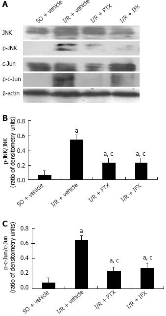
Figure 6 Tumor necrosis factor-α mediated c-Jun N-terminal kinase activation response to mucosal injury in ischemia-reperfusion-induced intestine.
Equal quantities of protein were subjected to Western blotting analysis, and β-actin was used as the control for loading. The results were expressed as a ratio of densitometry units. A: Western blotting analysis; B: The ratio of p-c-Jun N-terminal kinase (JNK) and JNK; C: The ratio of p-c-Jun and c-Jun. Values are mean ± SE. Six rats were tested in each group. aP < 0.05 vs sham-operation(SO) rats pretreated with vehicle (SO + vehicle); cP < 0.05 vs ischemia-reperfusion (I/R) rats pretreated with vehicle (I/R + vehicle). PTX: Pentoxifylline; IFX: Infliximab.
- Citation: Yang Q, Zheng FP, Zhan YS, Tao J, Tan SW, Liu HL, Wu B. Tumor necrosis factor-α mediates JNK activation response to intestinal ischemia-reperfusion injury. World J Gastroenterol 2013; 19(30): 4925-4934
- URL: https://www.wjgnet.com/1007-9327/full/v19/i30/4925.htm
- DOI: https://dx.doi.org/10.3748/wjg.v19.i30.4925


