Copyright
©2013 Baishideng Publishing Group Co.
World J Gastroenterol. Aug 14, 2013; 19(30): 4897-4906
Published online Aug 14, 2013. doi: 10.3748/wjg.v19.i30.4897
Published online Aug 14, 2013. doi: 10.3748/wjg.v19.i30.4897
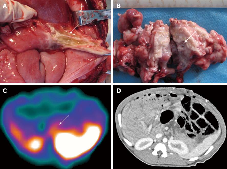
Figure 1 99mTc-ciprofloxacin scintigraphy has higher sensitivity and accuracy in the detection of bacterial infections than computed tomography (the swine came from group C).
A: The evidence of secondary infections in the peripancreatic fluid were confirmed by pathological examination and by bacterial smear or culture testing; B: Pancreatic necrosis was confirmed by pathological examination and by bacterial smear or culture testing; C: 99mTc-ciprofloxacin scintigraphy at 3 h demonstrated accumulation of radioactivity in the peripancreatic fluid and pancreatic necrosis (L/B = 3.37), indicating a true positive result; D: Enhanced computed tomography analysis showed low-density necrosis and peripancreatic fluid collection (arrow) without any indication of infection.
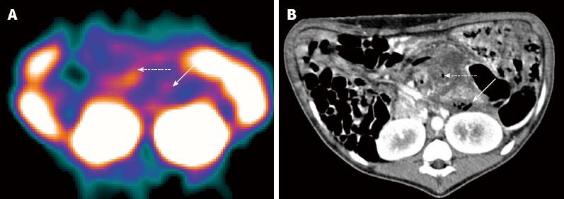
Figure 2 99mTc-ciprofloxacin and computed tomography imaging of a normal swine pancreas and both non-infected severe acute pancreatitis swine and an animal with secondary infection associated with severe acute pancreatitis.
A: No uptake indicating no infection, and a homogenous pancreatic parenchyma is evident in the normal pancreas by computed tomography imaging; B: A lower accumulation of 99mTc-ciprofloxacin in the pancreatic necroses of non-infected severe acute pancreatitis (SAP) swine; C: A higher accumulation of this agent in SAP swine with a secondary infection focus is evident.
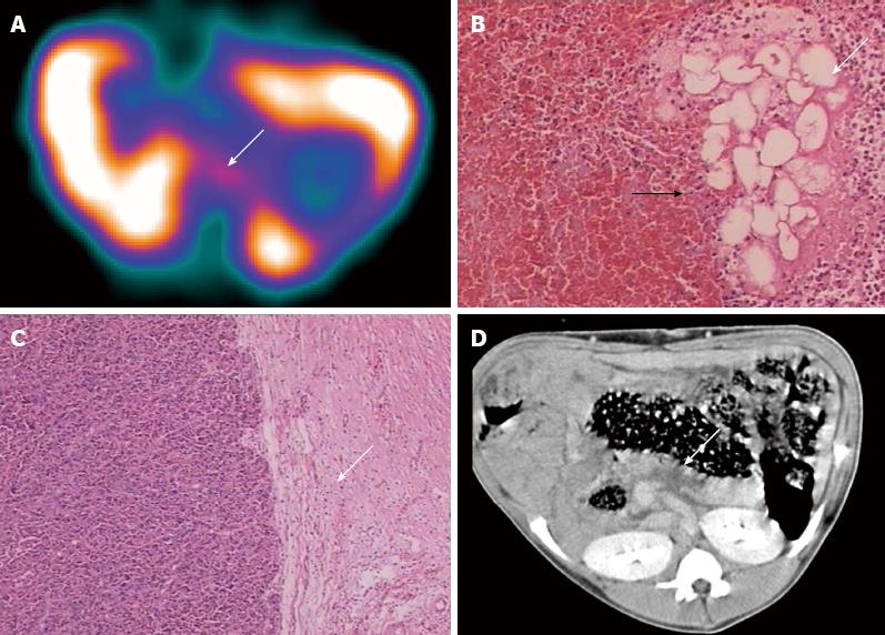
Figure 3 False-positive case of 99mTc-ciprofloxacin scintigraphy (the swine came from group B).
A: 99mTc-ciprofloxacin scintigraphy (arrow) is indicative of secondary infection (L/B = 2.15); B: However, light microscopy analysis showed pancreatic tissue and fat tissue necrosis (arrow), surrounding a marked hyperplastic area in granulated tissue (black arrow); C: Fibrous tissue (arrow), but no associated bacterial infection; D: Computed tomography image showed focal hypoattenuated areas within the pancreatic parenchyma (arrow) without gas bubbles.
Figure 4 Swine with severe acute pancreatitis and secondary bacterial infection (from group C).
A: The highest level of radioactivity accumulation was found by 99mTc-ciprofloxacin scintigraphy at 3 h (L/B = 3.42); B: Multiple bubbles scattered in the necrotic area and peripancreatic fluid were demonstrated on computed tomography images. Intestinal perforation caused by severe acute pancreatitis was found at autopsy.
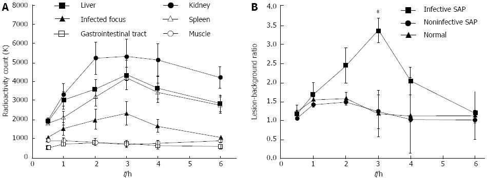
Figure 5 99mTc-ciprofloxacin scintigraphy results at different time points.
A: Curves showing radioactivity count changes over time in the abdominal tissues of swine with infected severe acute pancreatitis (SAP) are shown. These curves show high uptake in the kidneys and moderate uptake in the liver and spleen. No activity was observed in the areas of the muscle or gastrointestinal tract. The radioactive uptake by infectious foci increased gradually over time, reached a peak at 3 h, and then gradually decayed; B: Lesion-background ratio change curves for the normal pancreas, and non-infected and infected SAP pancreas in the swine model are shown. The curves show that the optimal lesion-background ratio occurred at 3 h after administration of Infecton in positive SAP animals.
- Citation: Wang JH, Sun GF, Zhang J, Shao CW, Zuo CJ, Hao J, Zheng JM, Feng XY. Infective severe acute pancreatitis: A comparison of 99mTc-ciprofloxacin scintigraphy and computed tomography. World J Gastroenterol 2013; 19(30): 4897-4906
- URL: https://www.wjgnet.com/1007-9327/full/v19/i30/4897.htm
- DOI: https://dx.doi.org/10.3748/wjg.v19.i30.4897


