
















©2013 Baishideng Publishing Group Co.
World J Gastroenterol. Mar 28, 2013; 19(12): 1919-1929
Published online Mar 28, 2013. doi: 10.3748/wjg.v19.i12.1919
Published online Mar 28, 2013. doi: 10.3748/wjg.v19.i12.1919
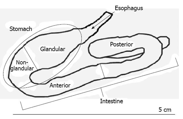
Figure 1 Anatomical diagram of the digestive tract of Oligosarcus hepsetus.
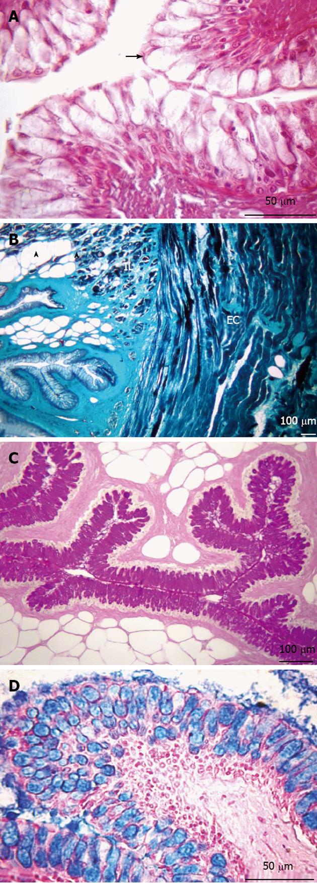
Figure 2 Transversal sections of the esophagus.
A: Non-keratinized stratified squamous epithelium (arrow). Hematoxylin and eosin stain; B: Presence of adipose tissue (arrowheads) in the submucosa layer. Muscular layer formed by two sub-layers, internal longitudinal (IL) and external circular (EC). Gomori’s trichrome stain; C: Presence of neutral glycoconjugates (GCs). Periodic acid-Schiff stain; D: Acid GCs. Alcian blue stain.
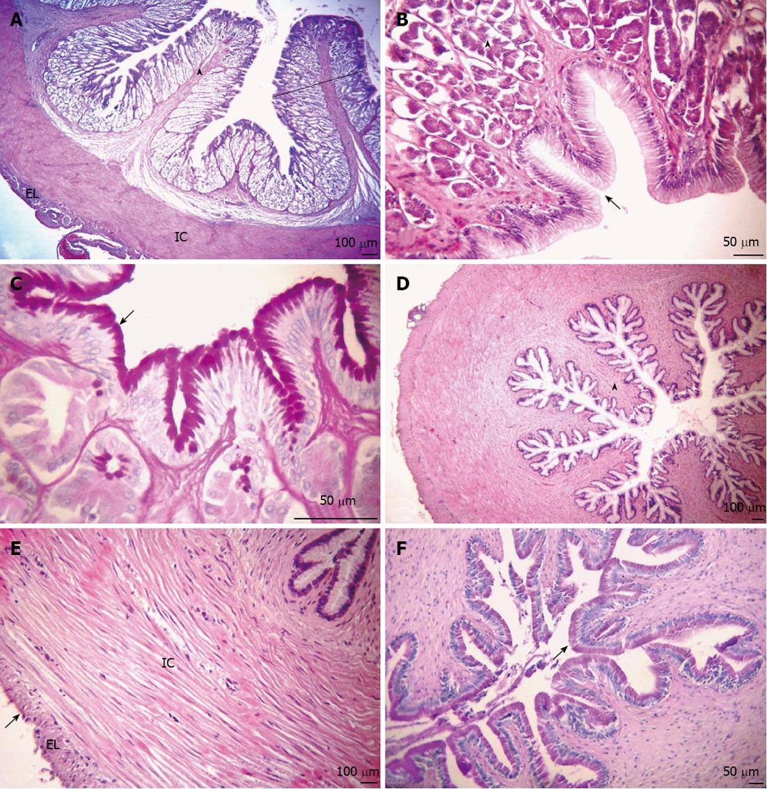
Figure 3 Transversal sections of the stomach.
A: Mucosa layer with many high gastric folds (arrowhead), thickening of the lamina propria caused by a large number of tubular gastric glands (outline). Organization of the internal circular (IC) and external longitudinal (EL) muscular layers. Hematoxylin and eosin (HE) stain; B: The simple columnar epithelium with mucus-secreting cells forming faveola gastrica (arrow) and fundic glands composed of oxynticopeptic cells (arrowhead). HE stain; C: Presence of neutral glycoconjugates (GCs) (arrow). Periodic acid Schiff (PAS) stain; D: Mucosa layer, together with the sub-mucosa and internal muscular layers, forming large longitudinal folds (arrowhead). HE stain; E: The muscular layer of smooth muscle fibers, composed of IC and EL sub-layers and serosa (arrow). HE stain; F: Neutral GCs (arrow). PAS stain. (A-C: Glandular region; D-F: Non-glandular region).
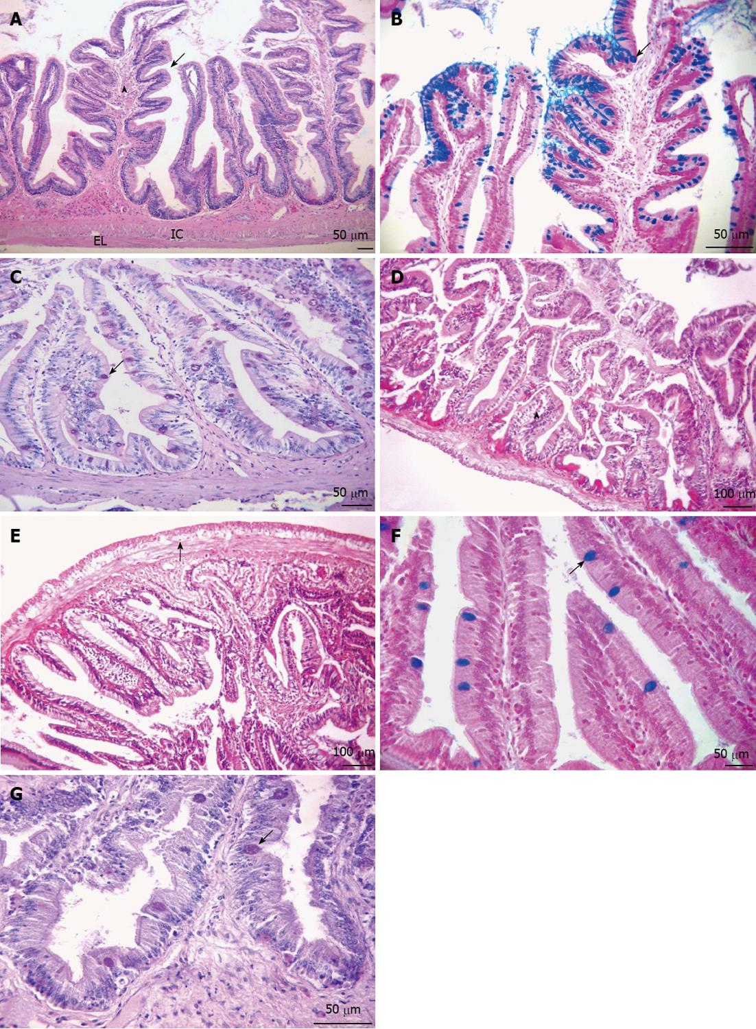
Figure 4 Longitudinal sections of the intestine.
A-C: Anterior portion; D-F: Posterior portion; A: Overview of the anterior intestine, showing the arrangement of small folds (arrowhead) presenting villi (arrow). Organization of the internal circular (IC) and external longitudinal (EL) muscular layers. Hematoxylin and eosin (HE) stain; B: Simple columnar epithelium with brush border and goblet cells, indicating the presence of acid glycoconjugates (GCs) (arrow). Alcian blue (AB) stain; C: Neutral GCs (arrows). Periodic acid Schiff (PAS) stain; D: Mucosa layer with thick folds (arrowhead). HE stain; E: Myenteric plexus (arrow). HE stain; F: Simple columnar epithelium with striated border and goblet cells with acid GCs (arrow). AB stain; G: Neutral GCs (arrow). PAS stain.
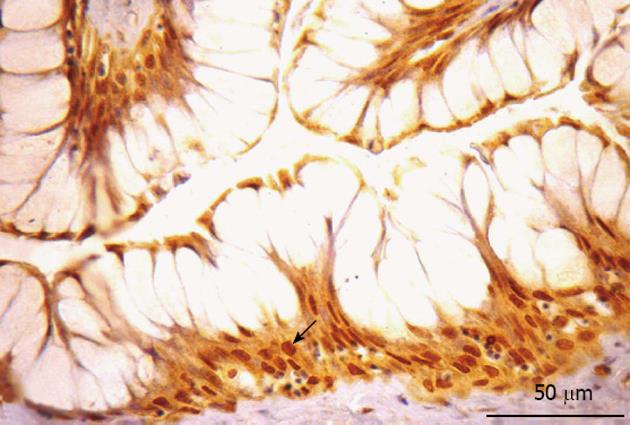
Figure 5 Somatostatin-immunorective cells in the esophagus.
Somatostatin-immunorective cell were detected in the basal layer of the stratified squamous epithelium (arrow).
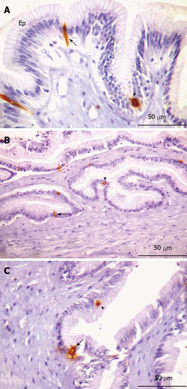
Figure 6 Serotonin-immunorective cells in the stomach.
A-B: Glandular region; C: Non-glandular region. A: Presence of open (arrow) and closed (arrowhead) type cells interspersed in the epithelium (Ep); B: Highlight of open (arrow) and closed (arrowhead) type immunorective (IR) cells; C: Indications of open (arrow) and closed (arrowhead) IR cells.
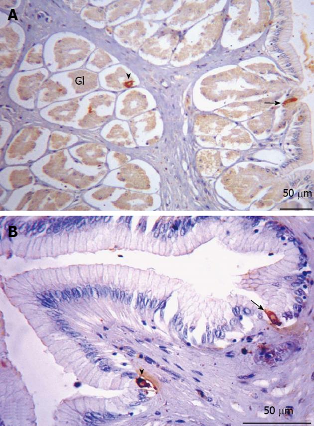
Figure 7 Somatostatin-immunorective cells in the stomach.
A: Glandular region. Immunorective (IR) cells along the entire region, of the open (arrow) and closed (arrowhead) types, present both in the lining epithelium and the glandular epithelium; B: Non-glandular region. Indications of IR cells of the open (arrow) and closed (arrowhead) types. Gl: Glandular.
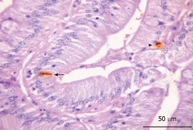
Figure 8 Cholecystokinin-immunorective cells in the posterior region of the intestine.
Epithelial marking of open (arrow) and closed (arrowhead) immunorective cells.
-
Citation: Vieira-Lopes DA, Pinheiro NL, Sales A, Ventura A, Araújo FG, Gomes ID, Nascimento AA. Immunohistochemical study of the digestive tract of
Oligosarcus hepsetus . World J Gastroenterol 2013; 19(12): 1919-1929 - URL: https://www.wjgnet.com/1007-9327/full/v19/i12/1919.htm
- DOI: https://dx.doi.org/10.3748/wjg.v19.i12.1919