©2012 Baishideng Publishing Group Co.
World J Gastroenterol. Oct 28, 2012; 18(40): 5799-5806
Published online Oct 28, 2012. doi: 10.3748/wjg.v18.i40.5799
Published online Oct 28, 2012. doi: 10.3748/wjg.v18.i40.5799
Figure 1 Macroscopic appearance of foregut neuroendocrine tumors in representative cases.
A: An esophageal neuroendocrine tumor (NET; case no. 16); B: A type I gastric NET (case no. 14); C: A type III gastric NET (case no. 17); D: A duodenal NET (case no. 11).
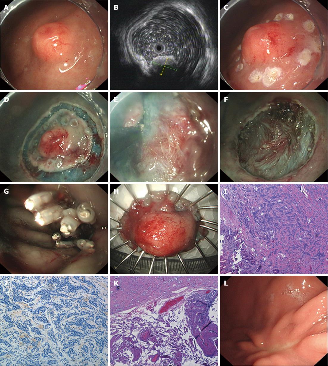
Figure 2 Endoscopic submucosal dissection of a type III gastric neuroendocrine tumor in a 71 year-old man (case no.
3). A: A submucosal tumor with a central depression of the gastric body; B: Endoscopic ultrasonography shows a mass invading the submucosa (maximum diameter, 18 mm); C-G: Processes of endoscopic submucosal dissection with the use of a soft transparent hood; H: Completely resected specimen; I: Microscopic examination of the completely resected specimen reveals a neuroendocrine tumor (NET) within the submucosa of the stomach (hematoxylin-eosin staining, × 40); J: The lesion was NET-G1 on the basis of proliferative activity (Ki-67 index < 2%, × 100); K: The basal margin was free of tumor invasion (hematoxylin-eosin staining, × 40); L: Endoscopic findings of scar 12 mo after endoscopic submucosal dissection.
- Citation: Li QL, Zhang YQ, Chen WF, Xu MD, Zhong YS, Ma LL, Qin WZ, Hu JW, Cai MY, Yao LQ, Zhou PH. Endoscopic submucosal dissection for foregut neuroendocrine tumors: An initial study. World J Gastroenterol 2012; 18(40): 5799-5806
- URL: https://www.wjgnet.com/1007-9327/full/v18/i40/5799.htm
- DOI: https://dx.doi.org/10.3748/wjg.v18.i40.5799


