©2012 Baishideng Publishing Group Co.
World J Gastroenterol. Oct 21, 2012; 18(39): 5649-5652
Published online Oct 21, 2012. doi: 10.3748/wjg.v18.i39.5649
Published online Oct 21, 2012. doi: 10.3748/wjg.v18.i39.5649
Figure 1 Gradual elevation of the right diaphragmatic border can be seen (arrows).
A: Chest radiography on admission; B: Two hours after admission; C: After thoracostomy.
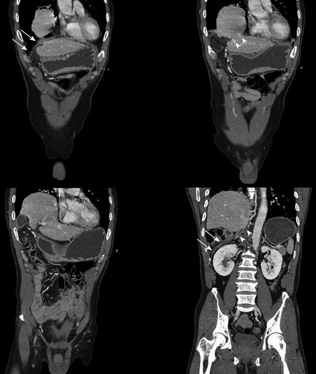
Figure 2 Chest computed tomography coronal reformatted images revealing intrathoracic displacement of the liver, bowel, and omentum through the defect (arrowheads) of the diaphragm (arrows).
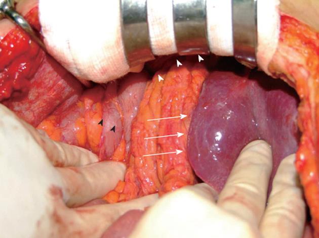
Figure 3 Herniation of the right hepatic lobe, gallbladder, transverse colon, and omentum through the diaphragmatic defect (white arrowheads), showing only the left hepatic lobe (white arrows) and residual transverse colon (black arrowheads) in the right upper abdomen.
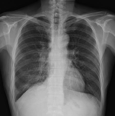
Figure 4 Chest radiograph 12 d after surgery showing normal positioning of the right diaphragmatic border compared to the preoperative chest radiograph.
- Citation: Baek SJ, Kim J, Lee SH. Hepatothorax due to a right diaphragmatic rupture related to duodenal ulcer perforation. World J Gastroenterol 2012; 18(39): 5649-5652
- URL: https://www.wjgnet.com/1007-9327/full/v18/i39/5649.htm
- DOI: https://dx.doi.org/10.3748/wjg.v18.i39.5649


