©2012 Baishideng Publishing Group Co.
World J Gastroenterol. Jul 28, 2012; 18(28): 3765-3769
Published online Jul 28, 2012. doi: 10.3748/wjg.v18.i28.3765
Published online Jul 28, 2012. doi: 10.3748/wjg.v18.i28.3765
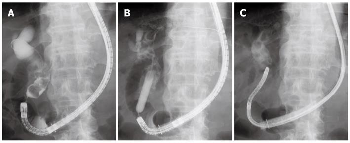
Figure 1 X-ray images.
A: Cholangiography revealed a dilated bile duct and multiple stones piled up at the lower to middle part of the bile duct; B: Papillary dilation using a large balloon catheter; C: An ultra-slim gastroscope was introduced directly into the bile duct.
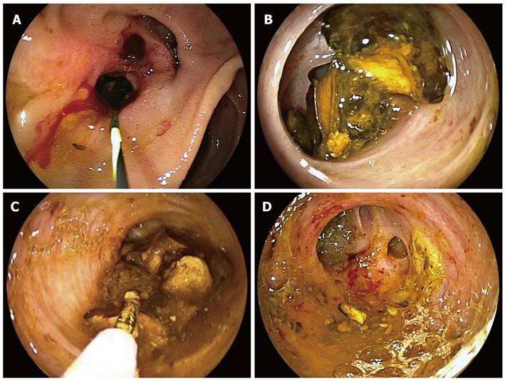
Figure 2 Endoscopic images.
A: The papilla of Vater after balloon dilation; B: Cholangioscopy showed multiple stones inside the bile duct; C, D: The stones were all removed with a 5-Fr basket catheter under direct visual control.
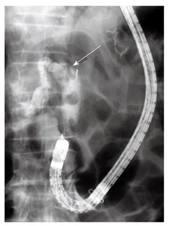
Figure 3 Cholangiography.
A smaller stone located distally was extracted with a basket catheter. Another stone was impacted inside the bile duct (arrow).
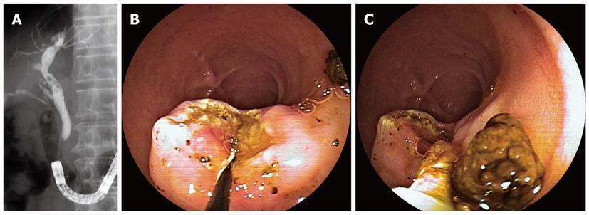
Figure 4 Direct cholangioscopy with an ultra-slim gastroscope.
A: Cholangioscopy revealed an impacted stone inside the bile duct; B: The stone was removed with a 5-Fr basket catheter under direct visual control; C: The proximal bile duct was also investigated.
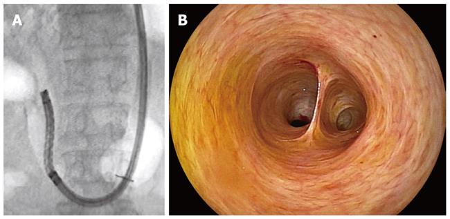
Figure 5 X-ray and endoscopic images.
A: Cholangiography revealed several small stones inside the bile duct; B: The papilla of Vater after endoscopic sphincterotomy; C: The bile duct stones were extracted with a basket catheter and a retrieval balloon.
Figure 6 Direct cholangioscopy.
A: An ultra-slim gastroscope was introduced directly into the bile duct after removal of bile duct stones; B: Cholangioscopy confirmed complete clearance of the duct.
- Citation: Koshitani T, Matsuda S, Takai K, Motoyoshi T, Nishikata M, Yamashita Y, Kirishima T, Yoshinami N, Shintani H, Yoshikawa T. Direct cholangioscopy combined with double-balloon enteroscope-assisted endoscopic retrograde cholangiopancreatography. World J Gastroenterol 2012; 18(28): 3765-3769
- URL: https://www.wjgnet.com/1007-9327/full/v18/i28/3765.htm
- DOI: https://dx.doi.org/10.3748/wjg.v18.i28.3765


