©2011 Baishideng Publishing Group Co.
World J Gastroenterol. Oct 28, 2011; 17(40): 4496-4502
Published online Oct 28, 2011. doi: 10.3748/wjg.v17.i40.4496
Published online Oct 28, 2011. doi: 10.3748/wjg.v17.i40.4496
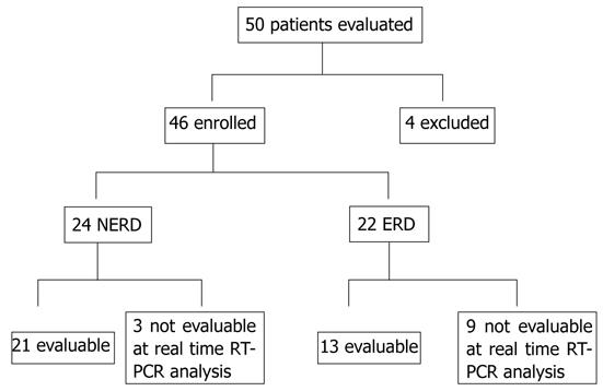
Figure 1 Study profile.
RT-PCR: Reverse transcription polymerase chain reaction; NERD: Non-erosive reflux disease; ERD: Erosive reflux disease.
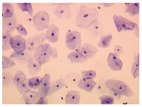
Figure 2 Cytological preparation from esophageal biopsy after epithelial cell isolation.
Only epithelial cells were present. Toluidine blue staining (300 ×).
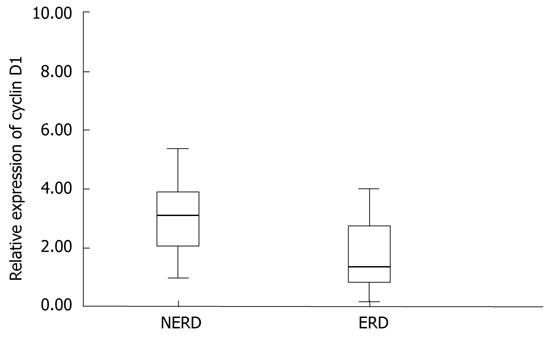
Figure 3 Box plots of relative expression of cyclin D1 mRNA by real-time RT-PCR analysis; median (bold line in box), and interquartile range (upper and lower lines of the box) in human esophageal mucosa of NERD and ERD patients (P < 0.
01). RT-PCR: Reverse transcription polymerase chain reaction; NERD: Non-erosive reflux disease; ERD: Erosive reflux disease.
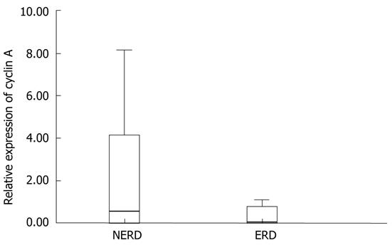
Figure 4 Box plots of relative expression of cyclin A mRNA by real-time RT-PCR analysis values; median (bold line in box), and interquartile range (upper and lower lines of the box) in human esophageal mucosa of NERD and ERD patients.
RT-PCR: Reverse transcription polymerase chain reaction; NERD: Non-erosive reflux disease; ERD: Erosive reflux disease.
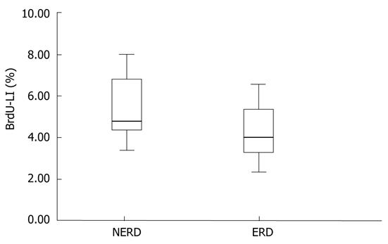
Figure 5 Box plots of BrdU-LI analysis values, median (bold line in the box), and interquartile range (upper and lower lines of the box) in human esophageal mucosa of NERD and ERD patients.
NERD: Non-erosive reflux disease; ERD: Erosive reflux disease.
- Citation: Calabrese C, Montanaro L, Liguori G, Brighenti E, Vici M, Gionchetti P, Rizzello F, Campieri M, Derenzini M, Trerè D. Cell proliferation of esophageal squamous epithelium in erosive and non-erosive reflux disease. World J Gastroenterol 2011; 17(40): 4496-4502
- URL: https://www.wjgnet.com/1007-9327/full/v17/i40/4496.htm
- DOI: https://dx.doi.org/10.3748/wjg.v17.i40.4496


