Copyright
©2011 Baishideng Publishing Group Co.
World J Gastroenterol. Aug 21, 2011; 17(31): 3591-3595
Published online Aug 21, 2011. doi: 10.3748/wjg.v17.i31.3591
Published online Aug 21, 2011. doi: 10.3748/wjg.v17.i31.3591
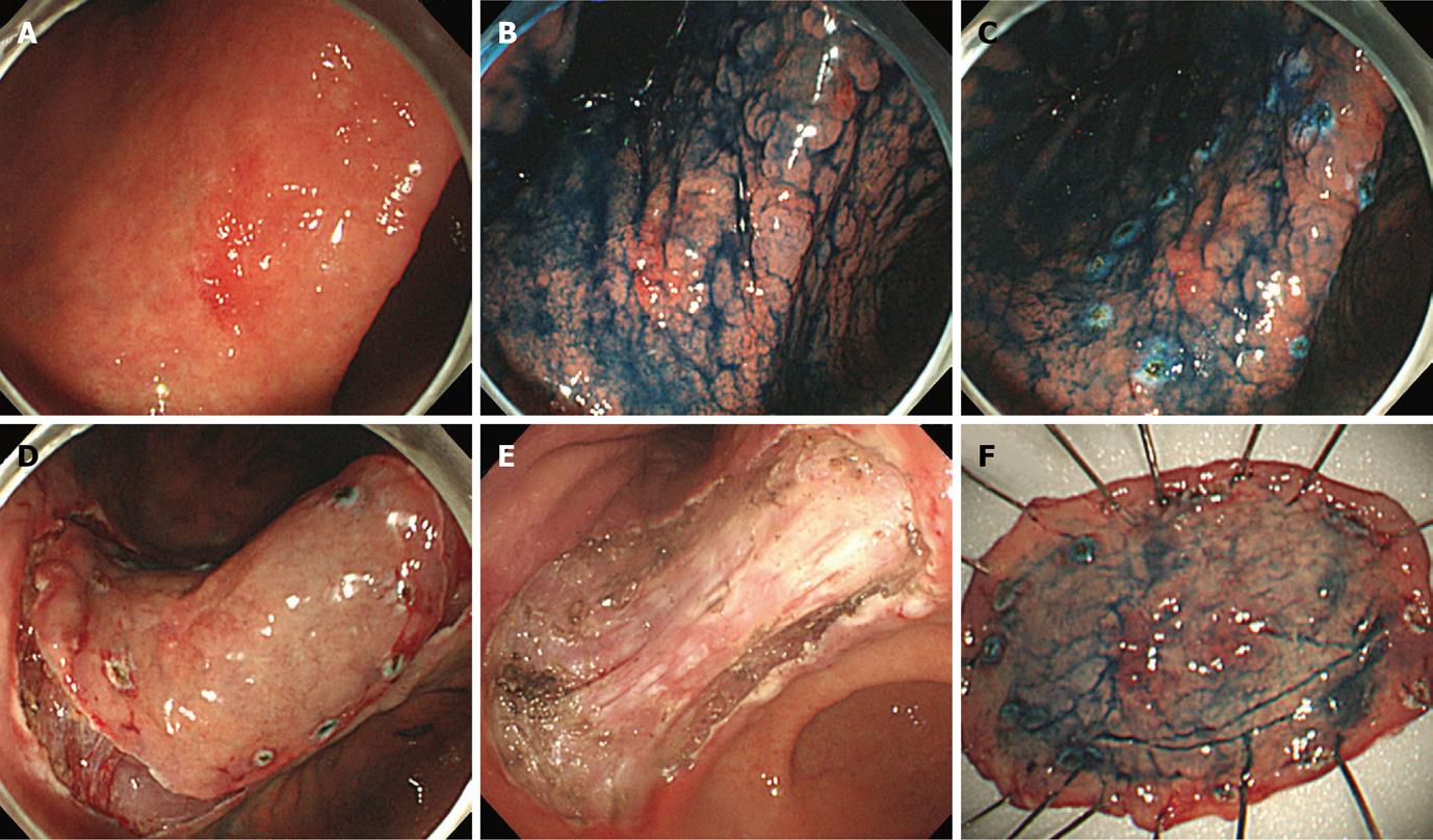
Figure 1 Endoscopic submucosal dissection procedure for early gastric cancer.
A: 1.5 cm × 1.2 cm sized hyperemic slightly elevated early gastric cancer was seen at the lesser curvature side of the lower body just above the gastric angle. Previous forceps biopsy results showed moderately differentiated adenocarcinoma; B: Indigo carmine dye was sprayed onto the lesion to define the lateral margin more clearly. Gastric mucosa around the cancer lesion showed severe metaplastic change; C: Using the tip of the needle knife, marking dots were made circumferentially at about 5 cm to 10 mm lateral to the estimated margin of the lesion; D: After submucosal injection of saline mixed with epinephrine and indigo carmine, a circumferential mucosal cutting was performed outside the marking dots to separate the lesion from the surrounding non-cancerous mucosa; E: After additional submucosal injection, direct dissection of the submucosal tissue was performed using an IT-knife and endoscopic hemostasis was carried out. A large artificial ulcer was made; F: The resected specimen with a central cancerous lesion. In the pathologic examination, a 1.8 cm × 1.1 cm sized moderately differentiated tubular adenocarcinoma limited in the mucosal layer was identified. The resection margin was free of cancer, and there was no lymphovascular invasion.
- Citation: Lee JH, Hong SJ, Jang JY, Kim SE, Seol SY. Outcome after endoscopic submucosal dissection for early gastric cancer in Korea. World J Gastroenterol 2011; 17(31): 3591-3595
- URL: https://www.wjgnet.com/1007-9327/full/v17/i31/3591.htm
- DOI: https://dx.doi.org/10.3748/wjg.v17.i31.3591


