©2011 Baishideng Publishing Group Co.
World J Gastroenterol. Aug 21, 2011; 17(31): 3580-3584
Published online Aug 21, 2011. doi: 10.3748/wjg.v17.i31.3580
Published online Aug 21, 2011. doi: 10.3748/wjg.v17.i31.3580
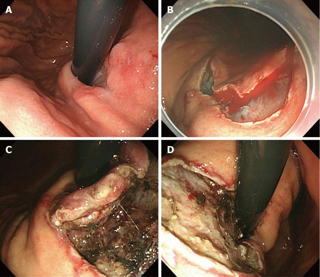
Figure 1 Lesion at the cardia, just below the gastroesophageal junction.
A: Retroflexed view of the cardia; B: A circumferential incision was made from the oral to the anal side, which is then vulnerable to bleeding; C: Submucosal dissection from the anal to the oral side; D: The lesion was completely resected.
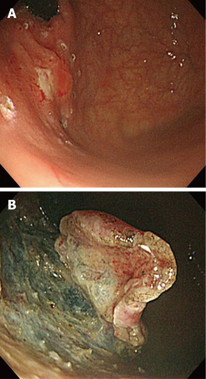
Figure 2 Lesion at the fundus.
A: The lesion was located between the fundus and the anterior side of the high body; B: Submucosal dissection was performed from the cardia to the fundus.
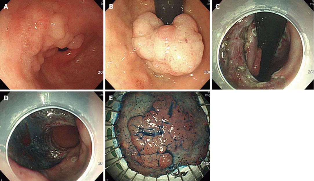
Figure 3 Lesion at the pyloric channel extending to the duodenal bulb.
A: Nodular elevated lesion involving the pyloric channel; B: Polypoid mass lesion at the duodenal bulb, retroflexed view; C: Incision and submucosal dissection were performed from the duodenal bulb to the antrum; D: The 180° circumferential dissection was completed; E: The en bloc resection was completed.
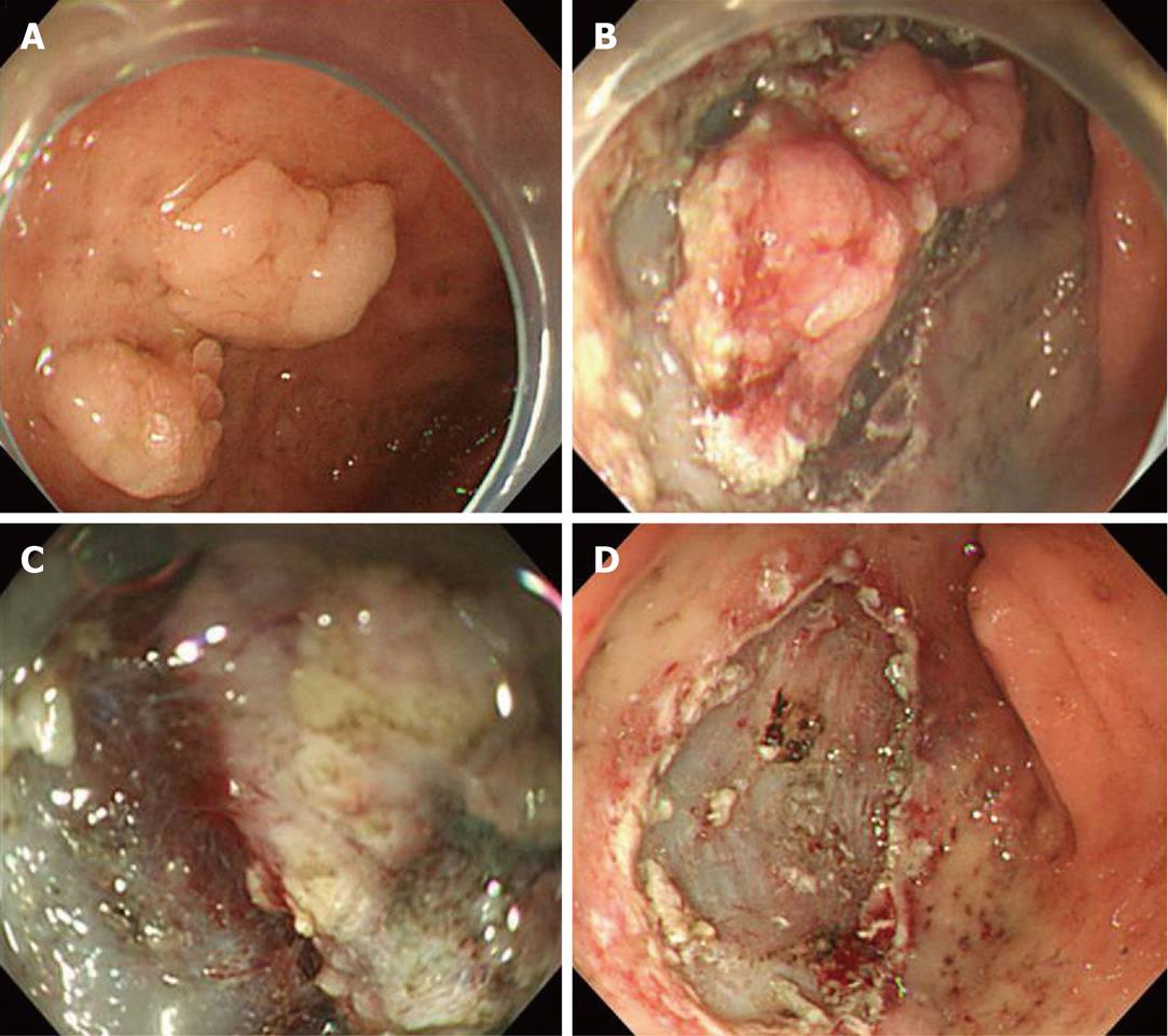
Figure 4 Lesion at the duodenum.
A: Two flat elevated lesions at the duodenal bulb; B: Circumferential incision; C: Submucosal dissection (the lifting of the lesion after submucosal injection was limited); D: The en bloc resection was completed.
- Citation: Kim KO, Kim SJ, Kim TH, Park JJ. Do you have what it takes for challenging endoscopic submucosal dissection cases? World J Gastroenterol 2011; 17(31): 3580-3584
- URL: https://www.wjgnet.com/1007-9327/full/v17/i31/3580.htm
- DOI: https://dx.doi.org/10.3748/wjg.v17.i31.3580


