Copyright
©2010 Baishideng Publishing Group Co.
World J Gastroenterol. Dec 21, 2010; 16(47): 6010-6015
Published online Dec 21, 2010. doi: 10.3748/wjg.v16.i47.6010
Published online Dec 21, 2010. doi: 10.3748/wjg.v16.i47.6010
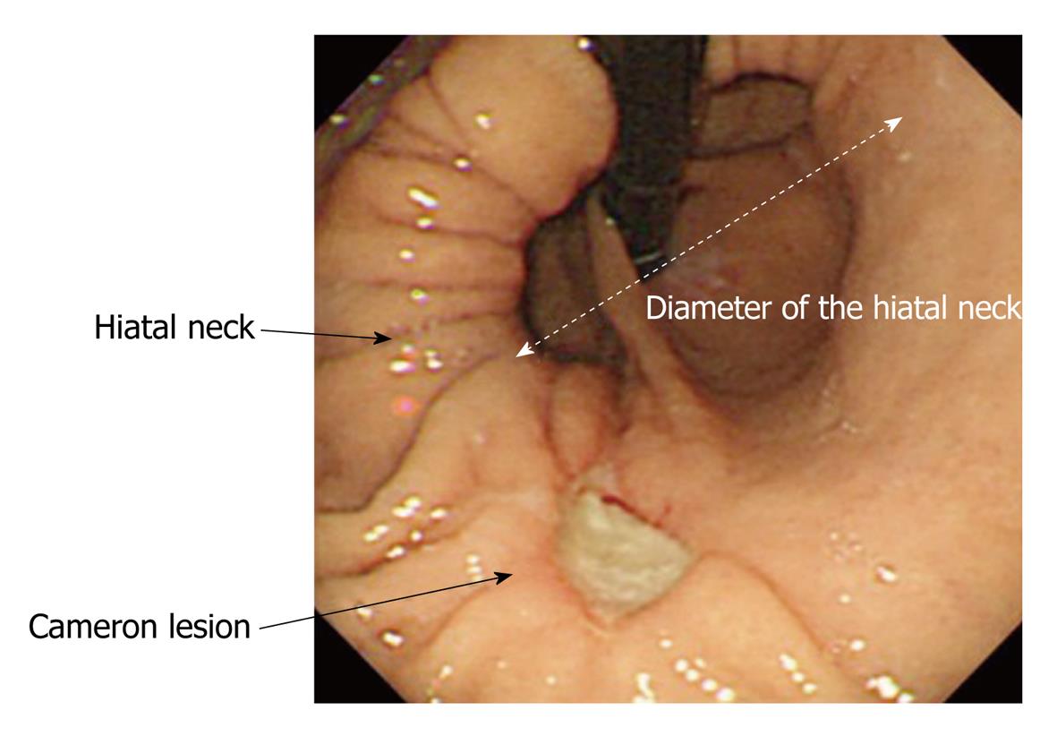
Figure 1 Representative image of the retroflex view of the gastric cardia in a patient with a large hiatal hernia and Cameron lesions.
Figure 2 Representative endoscopic images of an intact gastroesophageal flap valve (A) and an impaired gastroesophageal flap valve (B, C).
A loose and dull fold was classified as an impaired gastroesophageal flap valve (arrow in B).
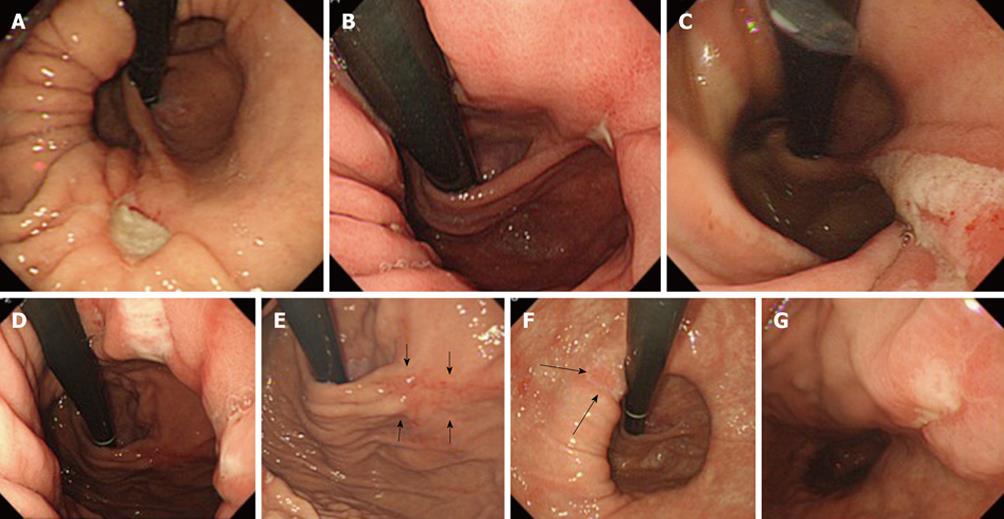
Figure 3 Representative endoscopic images of five patients with Cameron lesions.
A: Case #1 (no nonsteroidal anti-inflammatory drug, Hb5.4); B: Case #2 (loxoprofen + low dose aspirin, Hb5.3); C: Case #3 (diclofenac, Hb6.3); D, E: Case #4 (low dose aspirin, Hb8.0); F, G: Case #5 (low dose aspirin, Hb10.3). An ulceration scar was noted at the point at which the gastroesophageal flap valve fold diverged from the anterior stomach wall (arrows in E and F).
- Citation: Kaneyama H, Kaise M, Arakawa H, Arai Y, Kanazawa K, Tajiri H. Gastroesophageal flap valve status distinguishes clinical phenotypes of large hiatal hernia. World J Gastroenterol 2010; 16(47): 6010-6015
- URL: https://www.wjgnet.com/1007-9327/full/v16/i47/6010.htm
- DOI: https://dx.doi.org/10.3748/wjg.v16.i47.6010


