Copyright
©2010 Baishideng.
World J Gastroenterol. Jul 14, 2010; 16(26): 3299-3304
Published online Jul 14, 2010. doi: 10.3748/wjg.v16.i26.3299
Published online Jul 14, 2010. doi: 10.3748/wjg.v16.i26.3299
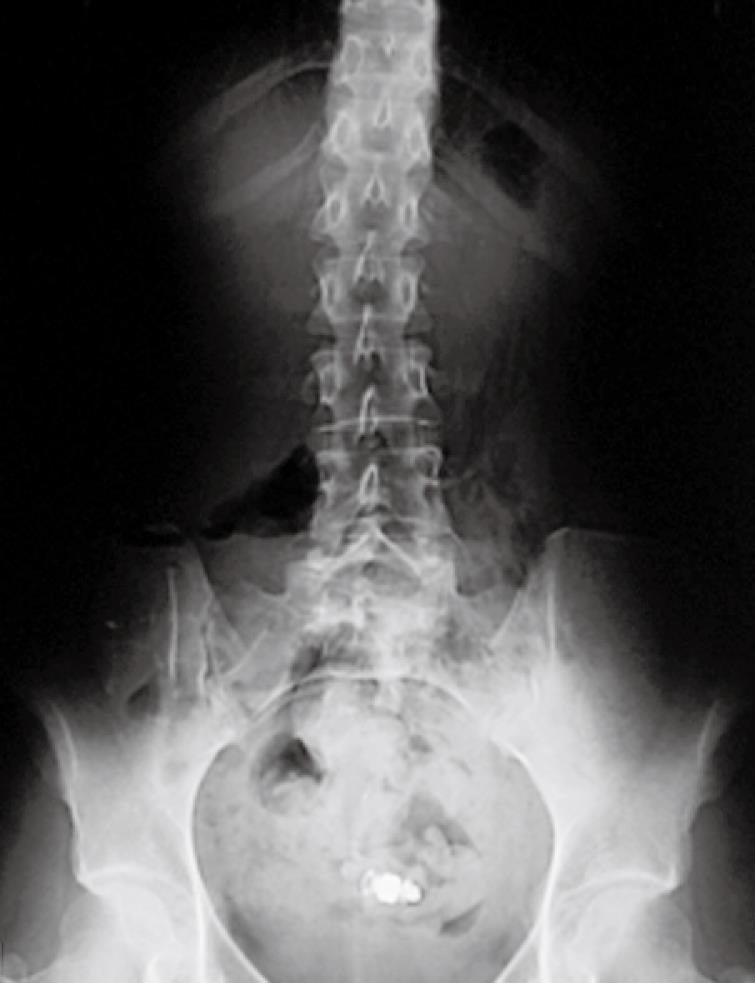
Figure 1 Plain film of the abdomen from one Crohn’s disease patient showing capsule retention for 12 wk, with no associated symptoms.
The patient had 2 anastomoses (ileo-ileal and ileo-colonic). Surgical removal was required, showing wireless capsule endoscopy within the “cul de sac” of the side-to-side ileo-ileal anastomosis not reachable by the endoscope.
Figure 2 Upper small bowel lesions detected at wireless capsule endoscopy in 3 patients with an established diagnosis of Crohn’s disease involving the distal ileum.
Aphthoid ulcer (A), deep ulcer (B) and one ulcerated stricture easily identified by wireless capsule endoscopy (C). All lesions compatible with Crohn’s disease detected by wireless capsule endoscopy appeared discontinuous and surrounded by macroscopically uninvolved mucosa.
- Citation: Petruzziello C, Onali S, Calabrese E, Zorzi F, Ascolani M, Condino G, Lolli E, Naccarato P, Pallone F, Biancone L. Wireless capsule endoscopy and proximal small bowel lesions in Crohn’s disease. World J Gastroenterol 2010; 16(26): 3299-3304
- URL: https://www.wjgnet.com/1007-9327/full/v16/i26/3299.htm
- DOI: https://dx.doi.org/10.3748/wjg.v16.i26.3299


