©2009 The WJG Press and Baishideng.
World J Gastroenterol. Nov 14, 2009; 15(42): 5266-5273
Published online Nov 14, 2009. doi: 10.3748/wjg.15.5266
Published online Nov 14, 2009. doi: 10.3748/wjg.15.5266
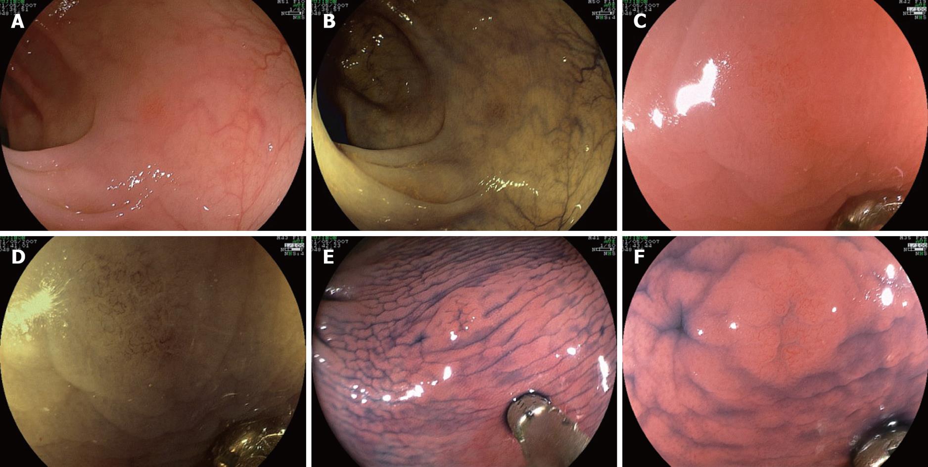
Figure 1 Presentation of a sequence of images for the different diagnostic modalities corresponding to a case included in study two (histopathological diagnosis: tubular adenoma with mild dysplasia).
A: Circumscribed erythema with loss of vascular pattern, which can be difficult to detect; B: Observation with Fujinon intelligent chromoendoscopy (FICE) 4. Note that the innominate grooves can be seen in the normal surrounding mucosa; C: Observation under 0.5% indigo carmine (IC), which reveals a 0-IIc depressed lesion 3 mm in size; D: High magnification view, showing vascular pattern type Sano II; E: Same magnification image observed under FICE 4, which provides enhanced vascular contrast; F: Under 0.5% IC plus magnification, the depression and the vascular patterns are evident.
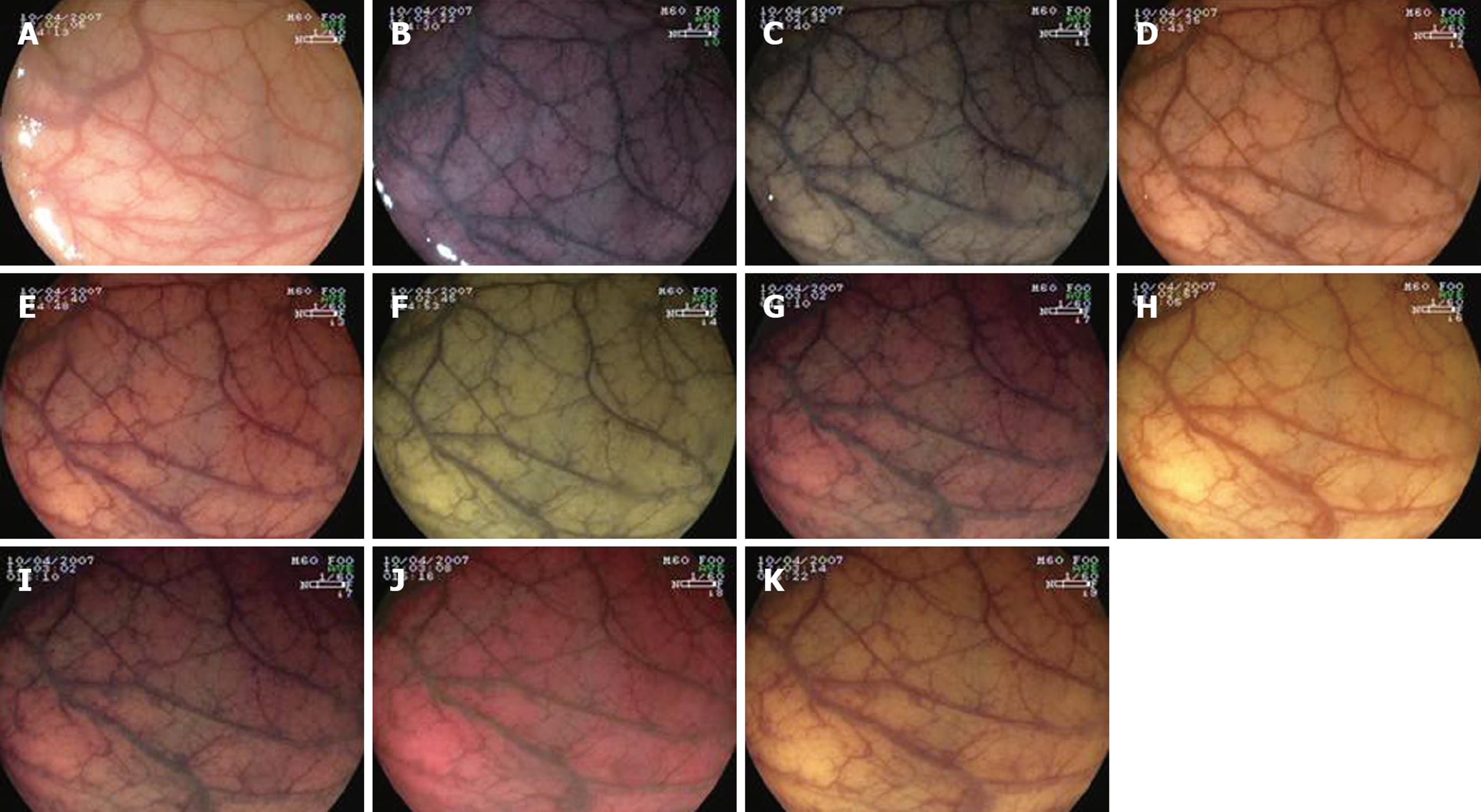
Figure 2 Images corresponding to the first study examining the evaluation of the vascular pattern without magnification.
A: Plain endoscopy; B: Filter 0; C: Filter 1; D: Filter 2; E: Filter 3; F: Filter 4; G: Filter 5; H: Filter 6; I: Filter 7; J: Filter 8; K: Filter 9.
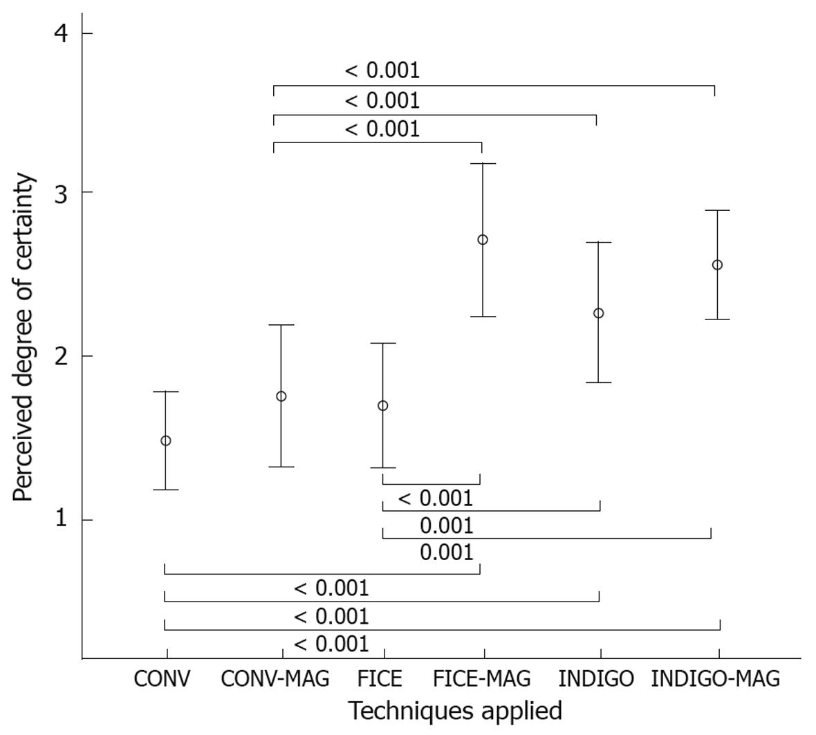
Figure 3 Degree of certainty in the endoscopic diagnosis for all participating endoscopists with the six diagnostic modalities employed, when endoscopic images were sequentially shown for each case (mean ± SD).
INDIGO-MAG: Indigo carmine plus magnification; FICE-MAG: FICE plus magnification; CONV-MAG: Conventional imaging plus magnification; CONV: Conventional.
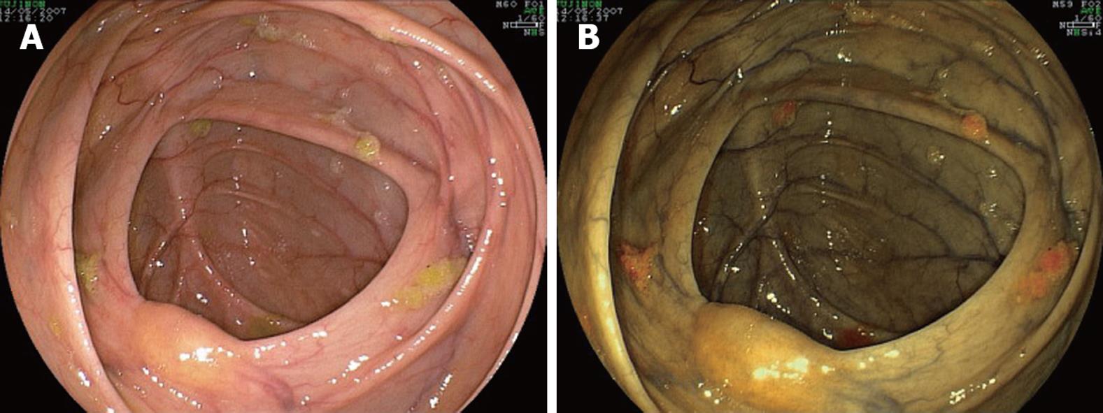
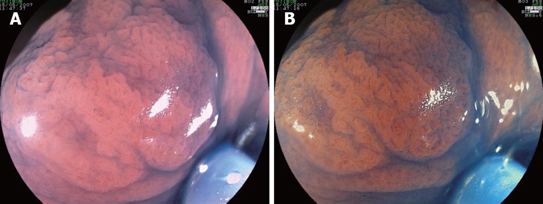
Figure 4 Observation of the colonic mucosa in the presence of remaining stool particles.
A: Plain endoscopy; B: FICE 4.
Figure 5 Sequence of images shown for the evaluation of mucosal contrast, in a 0-IIa flat elevated lesion, 3 mm in maximum diameter (histopathological diagnosis: tubular adenoma with mild dysplasia).
A: Plain endoscopy plus magnification; B: FICE 4 plus magnification; C: 0.5% IC plus magnification.
Figure 6 A flat elevated tubular adenoma, shown for the evaluation of FICE 6 plus IC.
A: 0.5% IC plus magnification; B: 0.5% IC plus magnification plus FICE 6.
- Citation: Parra-Blanco A, Jiménez A, Rembacken B, González N, Nicolás-Pérez D, Gimeno-García AZ, Carrillo-Palau M, Matsuda T, Quintero E. Validation of Fujinon intelligent chromoendoscopy with high definition endoscopes in colonoscopy. World J Gastroenterol 2009; 15(42): 5266-5273
- URL: https://www.wjgnet.com/1007-9327/full/v15/i42/5266.htm
- DOI: https://dx.doi.org/10.3748/wjg.15.5266


