©2009 The WJG Press and Baishideng.
World J Gastroenterol. Sep 21, 2009; 15(35): 4446-4448
Published online Sep 21, 2009. doi: 10.3748/wjg.15.4446
Published online Sep 21, 2009. doi: 10.3748/wjg.15.4446
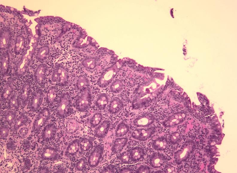
Figure 1 Antemortem biopsy from duodenum showed features of untreated celiac disease, despite a gluten-free diet.
Although later postmortem PCRof this biopsy documented a monoclonal population of T cells, frank lymphoma was not detected.
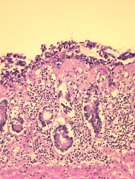
Figure 2 Postmortem small bowel section showed diffuse mucosal involvement with collagenous sprue.
Note eosin-stained sub-epithelial band. Trichrome staining was also positive (HE, × 20).
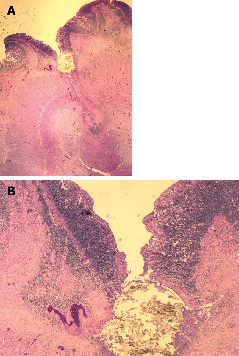
Figure 3 Autopsy showed ileum with a focal jejunal ulcer and perforation.
A: Low-power photomicrograph showed jejunal ulceration with perforation in an area of collagenous sprue in postmortem material; B: High-power photomicrograph showed perforated collagenous sprue.
- Citation: Freeman HJ, Webber DL. Free perforation of the small intestine in collagenous sprue. World J Gastroenterol 2009; 15(35): 4446-4448
- URL: https://www.wjgnet.com/1007-9327/full/v15/i35/4446.htm
- DOI: https://dx.doi.org/10.3748/wjg.15.4446


