©2009 The WJG Press and Baishideng.
World J Gastroenterol. Jun 14, 2009; 15(22): 2800-2804
Published online Jun 14, 2009. doi: 10.3748/wjg.15.2800
Published online Jun 14, 2009. doi: 10.3748/wjg.15.2800
Figure 1 Expression of semaphorin 5A and plexin B3 mRNA in 48 samples of primary gastric carcinoma tissues and its corresponding non-neoplastic mucosa as well as matched regional lymph node metastasis.
A: A representative result (left panel) and summary (right panel) of semaphorin 5A and plexin B3 expression in 48 samples of primary gastric carcinoma (Ca) and its corresponding nonneoplastic mucosa (M) as well as matched regional lymph node metastasis (L) examined by RT-PCR. The expression of β-actin was used as an internal control; B: Real time RT-PCR for relative expression levels of semaphorin 5A and plexin B3 in 48 samples of primary gastric carcinoma (Ca) and its corresponding nonneoplastic mucosa (M) as well as matched regional lymph node metastasis (L).
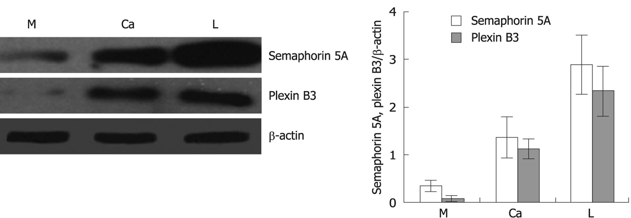
Figure 2 A representative result (left panel) and summary (right panel) of semaphorin 5A and plexin B3 protein expression in 48 samples of primary gastric carcinoma (Ca) and its corresponding nonneoplastic mucosa (M) as well as matched regional lymph node metastasis (L) examined by Western blotting.
The expression of β-actin was used as an internal control.
- Citation: Pan GQ, Ren HZ, Zhang SF, Wang XM, Wen JF. Expression of semaphorin 5A and its receptor plexin B3 contributes to invasion and metastasis of gastric carcinoma. World J Gastroenterol 2009; 15(22): 2800-2804
- URL: https://www.wjgnet.com/1007-9327/full/v15/i22/2800.htm
- DOI: https://dx.doi.org/10.3748/wjg.15.2800


