©2008 The WJG Press and Baishideng.
World J Gastroenterol. Aug 14, 2008; 14(30): 4810-4815
Published online Aug 14, 2008. doi: 10.3748/wjg.14.4810
Published online Aug 14, 2008. doi: 10.3748/wjg.14.4810
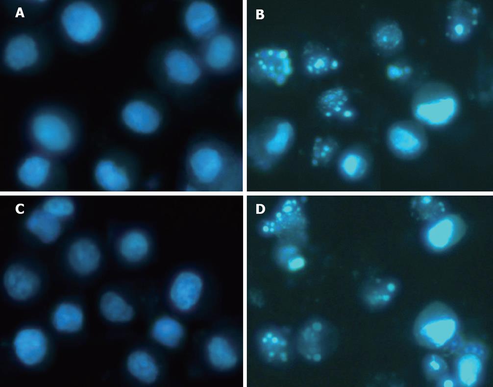
Figure 1 TSA (37.
5 ng/mL per 72 h in BGC-823 cells and 75 ng/mL per 72 h in SGC-7901 cells) induced apoptosis (Hoechst 33342 staining, × 400). A: No typical apoptotic BGC-823 cells were observed in control group; B: BGC-823 cells treated with TSA (37.5 ng/mL per 72 h) showed the typical apoptotic nuclear morphology; C: No typical apoptotic SGC-7901 cells were observed in control group; D: SGC-7901 cells treated with TSA (75 ng/mL for 72 h) showed the typical apoptotic nuclear morphology.
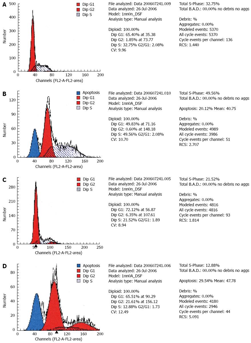
Figure 2 Effect of TSA on the cell cycle and apoptosis as analyzed by FACS.
A: Control of BGC-823 cells; B: Treatment with 37.5 ng/mL TSA for 72 h in BGC-823 cells; C: Control of SGC-7901 cells; D: Treatment with 75 ng/mL TSA for 72 h in SGC-7901 cells.
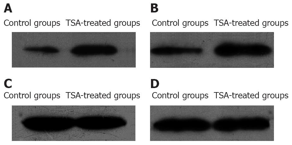
Figure 3 Cell lysates were analyzed by Western blots using anti-GAPDH and anti-acetyl-histone H3 antibody.
A: BGC-823 cells acetylated histone H3 (17 kDa); B: SGC-7901 cell acetylated histone H3 (17 kDa); C: BGC-823 cells GAPDH (loading control, 37 kDa); D: SGC-7901 cells GAPDH (loading control, 37 kDa).
- Citation: Zou XM, Li YL, Wang H, Cui W, Li XL, Fu SB, Jiang HC. Gastric cancer cell lines induced by trichostatin A. World J Gastroenterol 2008; 14(30): 4810-4815
- URL: https://www.wjgnet.com/1007-9327/full/v14/i30/4810.htm
- DOI: https://dx.doi.org/10.3748/wjg.14.4810


