©2008 The WJG Press and Baishideng.
World J Gastroenterol. Jul 14, 2008; 14(26): 4257-4259
Published online Jul 14, 2008. doi: 10.3748/wjg.14.4257
Published online Jul 14, 2008. doi: 10.3748/wjg.14.4257
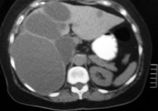
Figure 1 CT scan demonstrating multiple unilocular liver cysts.
The cysts are well delineated, smooth with uniform content, consistent with simple liver cysts.
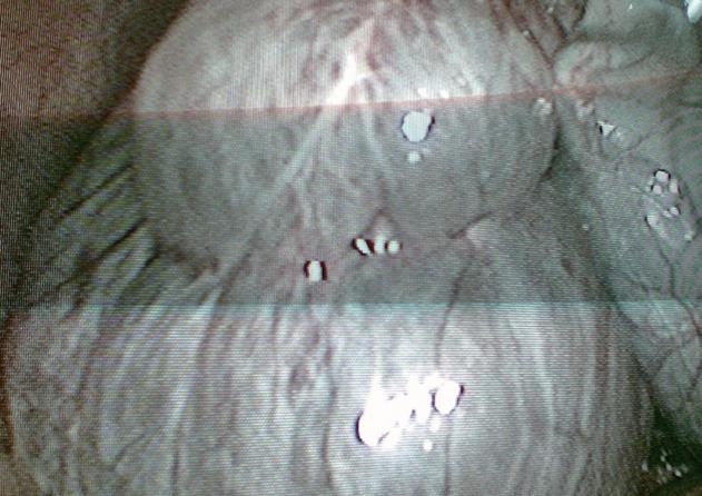
Figure 2 Laparoscopic view of the cysts and gallbladder.
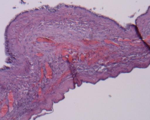
Figure 3 The cysts are lined with cuboidal to columnar non ciliated mucin secreting cells.
A moderate cellular stroma and an outer collagenous layer comprise the rest of the cyst wall (HE, × 40).
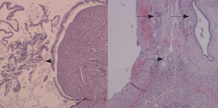
Figure 4 The wall of the largest cyst showing papillary projections of the lining epithelium (arrowhead), presence of microscopic cysts with the same epithelial lining (arrows), calcifications and hyaline degeneration of the stroma (HE, × 40).
- Citation: Manouras A, Lagoudianakis E, Alevizos L, Markogiannakis H, Kafiri G, Bramis C, Filis K, Toutouzas K. Laparoscopic fenestration of multiple giant biliary mucinous cystadenomas of the liver. World J Gastroenterol 2008; 14(26): 4257-4259
- URL: https://www.wjgnet.com/1007-9327/full/v14/i26/4257.htm
- DOI: https://dx.doi.org/10.3748/wjg.14.4257


