©2008 The WJG Press and Baishideng.
World J Gastroenterol. Apr 14, 2008; 14(14): 2174-2178
Published online Apr 14, 2008. doi: 10.3748/wjg.14.2174
Published online Apr 14, 2008. doi: 10.3748/wjg.14.2174
Figure 1 NGVE virus infected DEF cells analyzed by FACS, stained with annexin V-FITC/PI.
(A-C) display the results of the cells at 48, 96 and 144 h after NGVE virus infection. The proportion of non-apoptotic cells (c: Annexin V-FITC-/PI-), early apoptotic cells (d: Annexin V-FITC+/PI-), late apoptotic/necrotic cells (b: Annexin V-FITC+/PI+) and dead cells (a: Annexin V-FITC-/PI+).
Figure 2 Flow cytometry of apoptotic DEF cells as assessed by annexin V-FITC fluorescent intensity.
DEF cells are mock infected (A) and infected with NGVE virus (B). Cells harvested at 72 h p.i., and subsequently stained with annexin V-FITC/PI. One million cells are analyzed by flow cytometry, data are presented as fluorescent intensity units of annexin V-FITC (abscissa) and number of counts cells (ordinate). The M1 and M2 gates demarcate annexin V-FITC negative populations (non-apoptotic cells) and positive (apoptotic cells) populations.
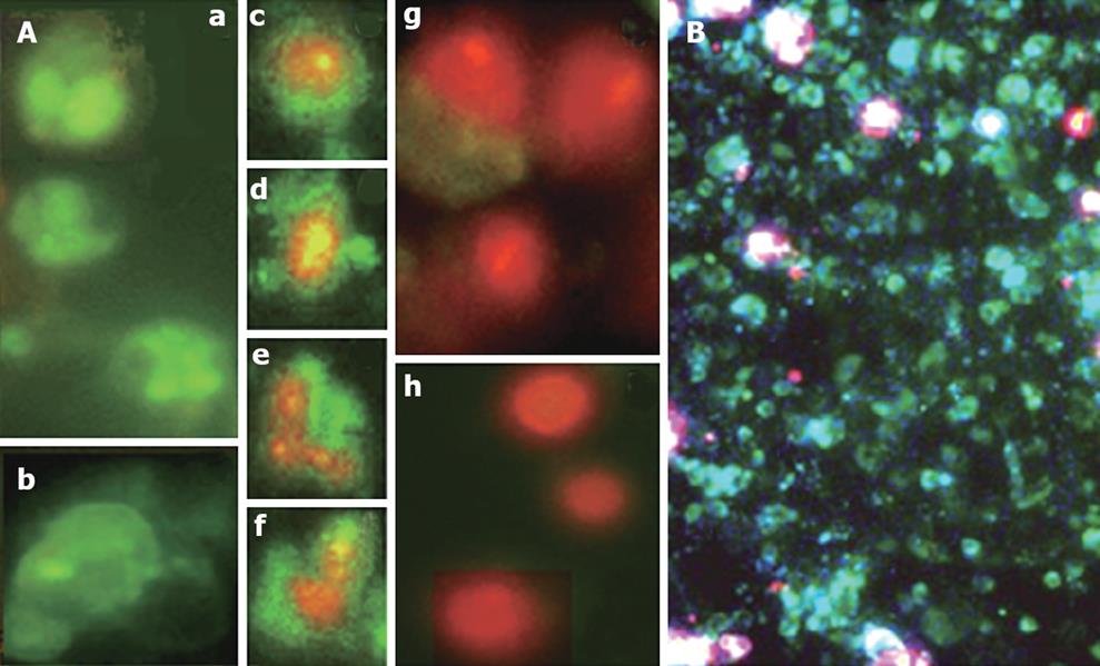
Figure 3 Apoptotic DEF cells induced by NGVE virus infection stained with Annexin V-FITC/PI and observed under fluorescence microscope.
The samples are analyzed for green fluorescence (FITC) and red fluorescence (PI). A: Different labeling patterns of the NGVE virus infected cells: early apoptotic cells, annexin V-FITC positive and PI negative (a and b); necrotic or late apoptotic cells, both annexin V-FITC and PI positive (c-f); dead cells, annexin V-FITC negative and PI positive (g and h); B: 72 h p.i., the early and late apoptotic cells.
-
Citation: Chen S, Cheng AC, Wang MS, Peng X. Detection of apoptosis induced by new type gosling viral enteritis virus
in vitro through fluorescein annexin V-FITC/PI double labeling. World J Gastroenterol 2008; 14(14): 2174-2178 - URL: https://www.wjgnet.com/1007-9327/full/v14/i14/2174.htm
- DOI: https://dx.doi.org/10.3748/wjg.14.2174


