©2008 The WJG Press and Baishideng.
World J Gastroenterol. Jan 7, 2008; 14(1): 129-131
Published online Jan 7, 2008. doi: 10.3748/wjg.14.129
Published online Jan 7, 2008. doi: 10.3748/wjg.14.129
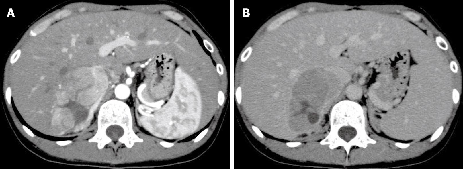
Figure 1 A: Dynamic enhanced CT showed a nodular lesion in the liver that was enhanced in the early image.
However, the interior of the mass showed a low-density area; B: In the subsequent late phase, the lesion revealed washout of contrast enhancement.
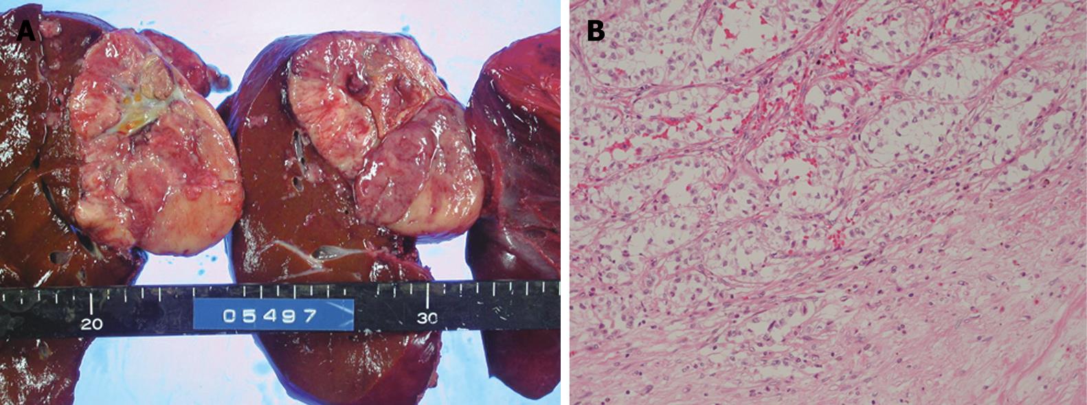
Figure 2 A: The tumor had a fibrous capsule.
Its cut surface was whitish, and tumorous tissue had a necrotic part at the center; B: Histological examination showed sheet-like growth of clear, atypical cells (HE, × 40).
- Citation: Takahashi A, Saito H, Kanno Y, Abe K, Yokokawa J, Irisawa A, Kenjo A, Saito T, Gotoh M, Ohira H. Case of clear-cell hepatocellular carcinoma that developed in the normal liver of a middle-aged woman. World J Gastroenterol 2008; 14(1): 129-131
- URL: https://www.wjgnet.com/1007-9327/full/v14/i1/129.htm
- DOI: https://dx.doi.org/10.3748/wjg.14.129


