Copyright
©2007 Baishideng Publishing Group Co.
World J Gastroenterol. Jan 21, 2007; 13(3): 467-469
Published online Jan 21, 2007. doi: 10.3748/wjg.v13.i3.467
Published online Jan 21, 2007. doi: 10.3748/wjg.v13.i3.467
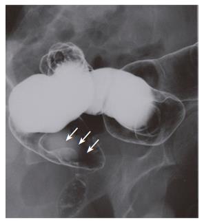
Figure 1 Double-contrast barium enema revealing a 20 mm × 15 mm sessile lesion in the periproctic area (arrows).
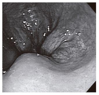
Figure 2 Colonoscopy disclosing a sessile lesion with a smooth surface.
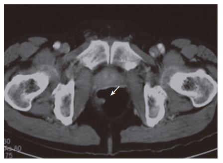
Figure 3 Enhanced CT of the lower abdomen demonstrating a fat-containing and slightly enhanced mass at a slice level corresponding to the right side of the lower rectum (arrow).
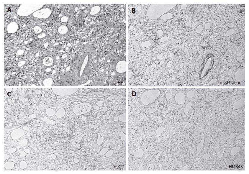
Figure 4 The tumor composed of a mixture of fat cells, proliferated blood vessels, spindle cells and immature mesenchymal cells (A), smooth muscle of a blood vessel demonstrating staining for alpha-smooth muscle actin but not for spindle cells (B), no tumor cells but only a few histiocytes displaying staining for c-kit (C), and no cells showing positive staining for HBM-45 (D).
- Citation: Ishizuka M, Nagata H, Takagi K, Horie T, Abe A, Kubota K. Rectal angiolipoma diagnosed after surgical resection: A case report. World J Gastroenterol 2007; 13(3): 467-469
- URL: https://www.wjgnet.com/1007-9327/full/v13/i3/467.htm
- DOI: https://dx.doi.org/10.3748/wjg.v13.i3.467


