Copyright
©2007 Baishideng Publishing Group Co.
World J Gastroenterol. Jul 21, 2007; 13(27): 3770-3772
Published online Jul 21, 2007. doi: 10.3748/wjg.v13.i27.3770
Published online Jul 21, 2007. doi: 10.3748/wjg.v13.i27.3770
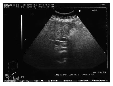
Figure 1 Transabdominal ultrasonography showing four parallel ducts.
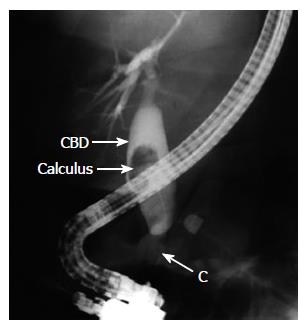
Figure 2 ERCP showing right CBD with calculus in its lumen (CBD: common bile duct; C:canula).
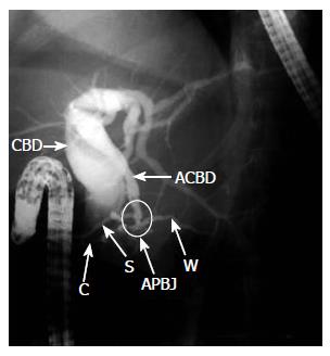
Figure 3 ERCP showing left CBD (CBD: common bile duct; ACBD: accessory common bile duct; APBJ: anomalous pancreaticobiliary junction; W: main pancreatic duct-Santorini; C: canula).
- Citation: Djuranovic SP, Ugljesic MB, Mijalkovic NS, Korneti VA, Kovacevic NV, Alempijevic TM, Radulovic SV, Tomic DV, Spuran MM. Double common bile duct: A case report. World J Gastroenterol 2007; 13(27): 3770-3772
- URL: https://www.wjgnet.com/1007-9327/full/v13/i27/3770.htm
- DOI: https://dx.doi.org/10.3748/wjg.v13.i27.3770


