Copyright
©2007 Baishideng Publishing Group Co.
World J Gastroenterol. Jul 21, 2007; 13(27): 3763-3764
Published online Jul 21, 2007. doi: 10.3748/wjg.v13.i27.3763
Published online Jul 21, 2007. doi: 10.3748/wjg.v13.i27.3763
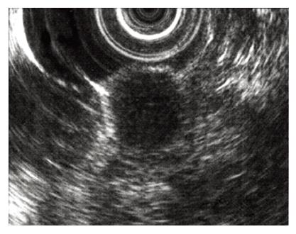
Figure 1 EUS revealed localized hypoechoic mass below the mucosal layer.
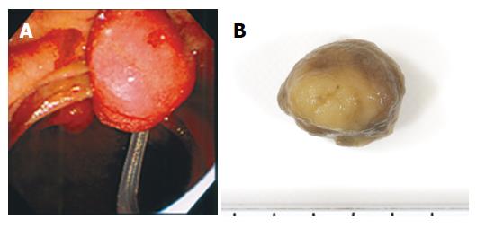
Figure 2 A: Endoscopic snare papillectomy of minor papilla was performed; B: Macroscopically, the tumor was yellowish and hard.
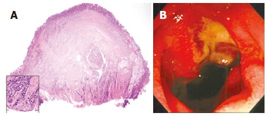
Figure 3 A: Histologically, the specimens showed invasive carcinoid tumor cells and the resected cut-end margin showed cancer cells; B: Duodenoscop revealed ulcer formation after the papillectomy.
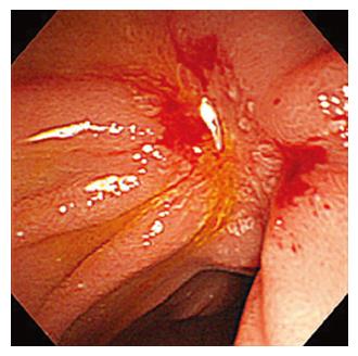
Figure 4 There was no evidence of recurrence 18 mo after the papillectomy.
- Citation: Itoi T, Sofuni A, Itokawa F, Tsuchiya T, Kurihara T, Moriyasu F. Endoscopic resection of carcinoid of the minor duodenal papilla. World J Gastroenterol 2007; 13(27): 3763-3764
- URL: https://www.wjgnet.com/1007-9327/full/v13/i27/3763.htm
- DOI: https://dx.doi.org/10.3748/wjg.v13.i27.3763


