Copyright
©2007 Baishideng Publishing Group Co.
World J Gastroenterol. Jul 21, 2007; 13(27): 3758-3759
Published online Jul 21, 2007. doi: 10.3748/wjg.v13.i27.3758
Published online Jul 21, 2007. doi: 10.3748/wjg.v13.i27.3758
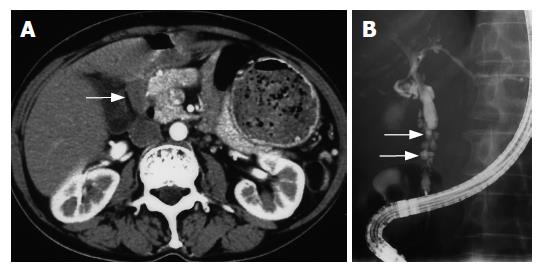
Figure 1 A: Abdominal computed tomography reveals markedly enlarged lymph nodes (arrow); B: Endoscopic retrograde cholangiography shows a bile duct stricture with a diverticulum-like outpouching (arrows).
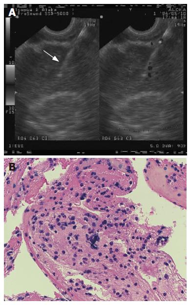
Figure 2 A: Endosonographic findings during EUS-FNA.
The arrow indicates the biopsy needle inside an enlarged lymph node; B: Histological features the specimen obtained by EUS-FNA.
- Citation: Tsukinaga S, Imazu H, Uchiyama Y, Kakutani H, Kuramoti A, Kato M, Kanazawa K, Kobayashi T, Searashi Y, Tajiri H. Diagnostic approach using endosonography guided fine needle aspiration for lymphadenopathy in primary sclerosing cholangitis. World J Gastroenterol 2007; 13(27): 3758-3759
- URL: https://www.wjgnet.com/1007-9327/full/v13/i27/3758.htm
- DOI: https://dx.doi.org/10.3748/wjg.v13.i27.3758


