©2007 Baishideng Publishing Group Inc.
World J Gastroenterol. Jul 14, 2007; 13(26): 3625-3630
Published online Jul 14, 2007. doi: 10.3748/wjg.v13.i26.3625
Published online Jul 14, 2007. doi: 10.3748/wjg.v13.i26.3625
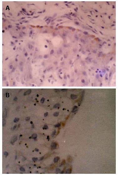
Figure 1 The IHC staining results of the placenta of HBV carrier mothers.
A: HBsAg positive cells in the vascular endothelial cell of the placenta villi (× 40); B: HBsAg positive cells in the trophoblastic cell of on the surface of placenta villi (× 40).
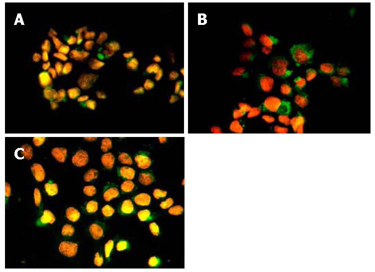
Figure 2 Immunofluorescence staining results of trophoblastic cells of different co-incubation time with HBV positive serum.
A: 8 h; B: 24 h; C: 48 h.
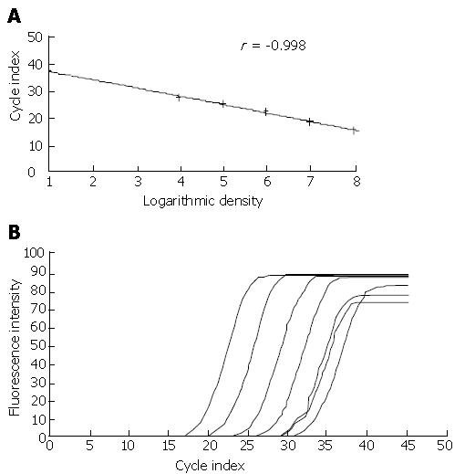
Figure 3 HBV DNA detection results in the trophoblastic cell by RT-PCR.
A: standard curve; B: detective result.
- Citation: Bai H, Zhang L, Ma L, Dou XG, Feng GH, Zhao GZ. Relationship of hepatitis B virus infection of placental barrier and hepatitis B virus intra-uterine transmission mechanism. World J Gastroenterol 2007; 13(26): 3625-3630
- URL: https://www.wjgnet.com/1007-9327/full/v13/i26/3625.htm
- DOI: https://dx.doi.org/10.3748/wjg.v13.i26.3625


