Copyright
©2007 Baishideng Publishing Group Co.
World J Gastroenterol. Apr 28, 2007; 13(16): 2363-2368
Published online Apr 28, 2007. doi: 10.3748/wjg.v13.i16.2363
Published online Apr 28, 2007. doi: 10.3748/wjg.v13.i16.2363
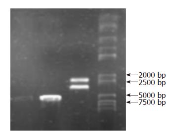
Figure 1 Gel electropho-resis: PBluescript-Human Selenoprotein P has 6.
4 kb, SeP segment about 2 kb and plasmid carrier about 4.4 kb when digested with SacI and XhoI.
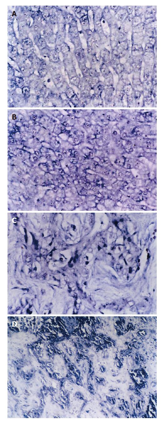
Figure 2 SePmRNA expressions in liver tissues.
A: In normal liver tissues, the positive signals were in nucleus and cytoplasm, mostly in nucleolus, and the stained granules are large in nucleolus and around nucleus (ISH × 400); B: In liver cirrhosis tissues, the positive signals were mainly in nucleolus and cytoplasma, and the signals around nucleus and inner nucleus were less than in normal liver tissues (ISH × 400); C: In HCC tissues, the positive signals were in the cytoplasm, but less in nucleus. (ISH × 400); D: In hepatic interstitial substance, the positive signals were in the matrix, mainly in vascular endothelial cells and lymphocytes of vasculature (ISH × 400).
- Citation: Li CL, Nan KJ, Tian T, Sui CG, Liu YF. Selenoprotein P mRNA expression in human hepatic tissues. World J Gastroenterol 2007; 13(16): 2363-2368
- URL: https://www.wjgnet.com/1007-9327/full/v13/i16/2363.htm
- DOI: https://dx.doi.org/10.3748/wjg.v13.i16.2363


