©2006 Baishideng Publishing Group Co.
World J Gastroenterol. Dec 28, 2006; 12(48): 7869-7873
Published online Dec 28, 2006. doi: 10.3748/wjg.v12.i48.7869
Published online Dec 28, 2006. doi: 10.3748/wjg.v12.i48.7869
Figure 1 Solitary mass type: Enhanced CT showing multiple enlarged lymph nodes fused into uneven density, huge lobular tu-mors with abdo-minal aorta and inferior vena cava encased and ascites.
Figure 2 Multi-ple-nodular type: Enhanced CT showing multiple, homogeneous density, enlarged lymph nodes in retroperitoneal region with as-cites.
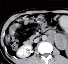
Figure 3 Multi-ple-nodular type: CT of portal ve-nous phase show-ing mesenteric multiple enlarged lymph nodes with superior mesen-teric artery en-cased.
Figure 4 Diff-use type: Enhanced CT showing diffuse, clear margins, homo-geneous density enlar-ged lymph nodes in the mesenteric and retroperitoneal region.
Figure 5 En-hanced CT show-ing multiple, homogeneous density, enlarged lymph nodes in para-abdominal aorta and multiple, low-density lesions in the spleen (splenic lymphoma).
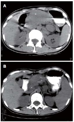
Figure 6 A: Plain CT showing a uniform density, slightly lobular tumor (formed by multiple enlarged lymph nodes) in the lesser omentum; B: The tumor having a notable shrinkage in size and flecked calcifications found within the tumor after treatment on plain CT.
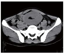
Figure 7 En-hanced CT show-ing notable cir-cular thickening of intestinal wall with homogeneous density.
- Citation: Yu RS, Zhang WM, Liu YQ. CT diagnosis of 52 patients with lymphoma in abdominal lymph nodes. World J Gastroenterol 2006; 12(48): 7869-7873
- URL: https://www.wjgnet.com/1007-9327/full/v12/i48/7869.htm
- DOI: https://dx.doi.org/10.3748/wjg.v12.i48.7869


