Copyright
©2006 Baishideng Publishing Group Co.
World J Gastroenterol. Nov 7, 2006; 12(41): 6727-6729
Published online Nov 7, 2006. doi: 10.3748/wjg.v12.i41.6727
Published online Nov 7, 2006. doi: 10.3748/wjg.v12.i41.6727
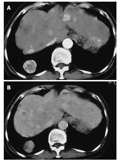
Figure 1 Abdominal incremental computed tomography (CT) (A) showing multiple contrast-enhanced tumors (arterial phase) and abdominal incremental CT scan (B) showing low density tumors (portal phase).
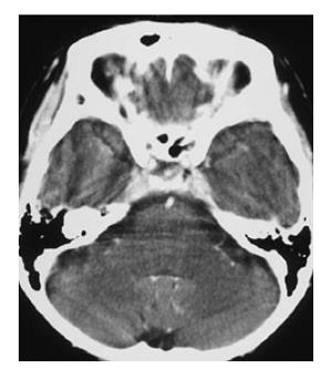
Figure 2 Brain contrast-enhanced CT image.
Abnormal enhancement around the sella turcica is observed.
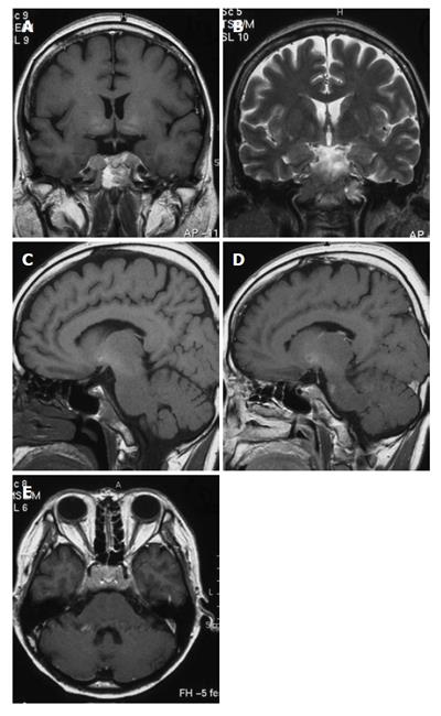
Figure 3 Brain MRI image.
A: Unenhanced coronal T1-weighted image reveals ill-defined mass around the sella turnica. The mass shows intermediate signal intensity; B: A mass reveals high signal intensity on coronal T2-weighted image; C: Unenhanced saggital T1-weighted image reveals ill defined mass involving the sella turcica. D, E: On contrast-enhanced saggital (D) and transverse (E) T1-weighted images, the mass shows inhomogeneous intermediate enhancement and involves the clivus, cavernous sinus and petrous apex. Meningeal thickening between the posterior sella turcica and the clivus and dilation of the superior carotid vein are observed.
- Citation: Kim SR, Kanda F, Kobessho H, Sugimoto K, Matsuoka T, Kudo M, Hayashi Y. Hepatocellular carcinoma metastasizing to the skull base involving multiple cranial nerves. World J Gastroenterol 2006; 12(41): 6727-6729
- URL: https://www.wjgnet.com/1007-9327/full/v12/i41/6727.htm
- DOI: https://dx.doi.org/10.3748/wjg.v12.i41.6727


