Copyright
©2006 Baishideng Publishing Group Co.
World J Gastroenterol. Jul 28, 2006; 12(28): 4593-4595
Published online Jul 28, 2006. doi: 10.3748/wjg.v12.i28.4593
Published online Jul 28, 2006. doi: 10.3748/wjg.v12.i28.4593
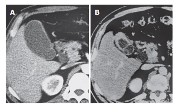
Figure 1 A: CT reveals diffuse thickening of the gallbladder wall and dilatation of the common bile duct to 14 mm; B: After 42 mo, CT reveals progressive thickening of the gallbladder wall, suspicious for gallbladder carcinoma.
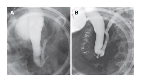
Figure 2 A: ERCP shows no pancreaticobiliary maljunction, because sphincter of Oddi affected pancreaticobiliary junction and connection between pancreatic and biliary ducts was not visible during the contraction phase of sphincter of Oddi; B: ERCP in the relaxation phase of sphincter of Oddi shows the length of the common channel to be 5 mm long.
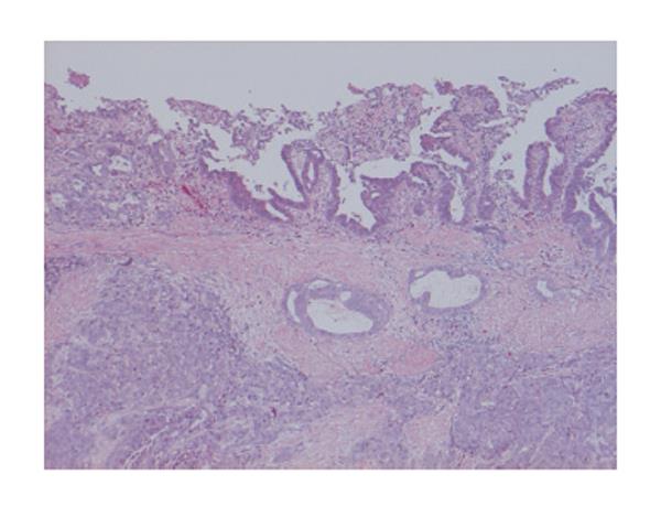
Figure 3 Histopathologic examination of the resected specimen demonstrates a moderately differentiated adenocarcioma of the gallbladder (HE stain, × 40).
- Citation: Sai JK, Suyama M, Kubokawa Y. A case of gallbladder carcinoma associated with pancreatobiliary reflux in the absence of a pancreaticobiliary maljunction: A hint for early diagnosis of gallbladder carcinoma. World J Gastroenterol 2006; 12(28): 4593-4595
- URL: https://www.wjgnet.com/1007-9327/full/v12/i28/4593.htm
- DOI: https://dx.doi.org/10.3748/wjg.v12.i28.4593


