©2006 Baishideng Publishing Group Co.
World J Gastroenterol. Jul 21, 2006; 12(27): 4435-4436
Published online Jul 21, 2006. doi: 10.3748/wjg.v12.i27.4435
Published online Jul 21, 2006. doi: 10.3748/wjg.v12.i27.4435
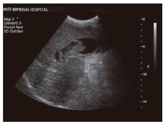
Figure 1 Ultra-sonography of the abdomen showing distended gall bladder containing large blood clot.
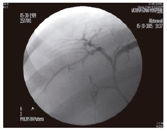
Figure 2 Intra-operative cholangiogram with normal appearing anatomy.
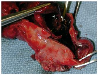
Figure 3 The opened specimen revealing a blood clot.
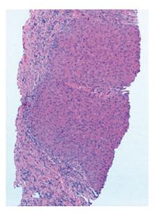
Figure 4 Hemtoxilin & eosin stain of the liver sampled from the case.
- Citation: Edden Y, Hilaire HS, Benkov K, Harris MT. Percutaneous liver biopsy complicated by hemobilia-associated acute cholecystitis. World J Gastroenterol 2006; 12(27): 4435-4436
- URL: https://www.wjgnet.com/1007-9327/full/v12/i27/4435.htm
- DOI: https://dx.doi.org/10.3748/wjg.v12.i27.4435


