Copyright
©2006 Baishideng Publishing Group Co.
World J Gastroenterol. May 28, 2006; 12(20): 3265-3270
Published online May 28, 2006. doi: 10.3748/wjg.v12.i20.3265
Published online May 28, 2006. doi: 10.3748/wjg.v12.i20.3265
Figure 1 HPD in a 48-year-old woman with cavernous hemangioma.
A: Pre- contrast administration transverse CT scan shows a large well-defined hypo-attenuating lesion located in segment II(curved open arrow); B: HAP CT scan shows a nodular enhanced hemangioma (curved open arrow) and wedge-shaped segmental homogeneous hyperattenuating areas (solid arrow) adjacent to the tumor; C: PVP CT scan shows the still nodular enhanced hemangioma (curved) and the peripheral area resumed isoattenuating.
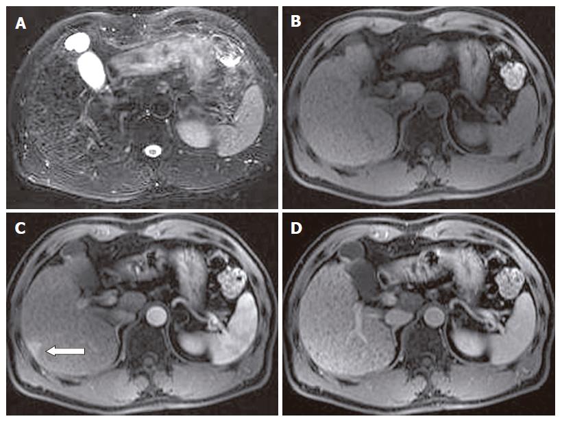
Figure 2 HPD in a 56-year-old man with liver cirrhosis.
A: Fat-saturated breath-hold turbo FSE T2-weighted transverse MR scan shows multiple punctuate hypointensity cirrhotic nodules and surrounding reticulated fibrotic liver parenchyma, without any obvious hyperintense tumoral lesions; B: pre- contrast administration transverse spoiled gradient-echo T1-weighted MR image (118/4.1; flip angle, 80°) shows no obvious hypointense tumoral lesions observed; C: transverse contrast-enhanced T1-weighted MR image obtained during HAP shows a subcapsular small patchy hyperintense tumor-like lesion (pseudolesion) (open arrow); D: transverse contrast-enhanced T1-weighted MR image obtained during PVP shows no lesion in that area.
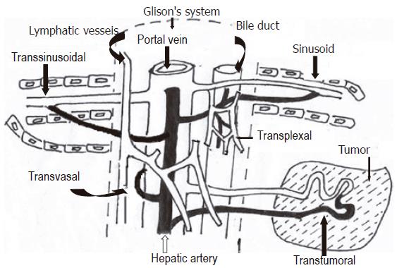
Figure 3 Schematic drawings showing hepatic microvasculature and arterioportal anastomoses or arterioportal shunts, including the transsinusoidal route (between the interlobular arterioles and portal venules or sinusoids) and the transplexal route (peribiliary plexus), transvasal route (perilymphatic vessels) and transtumoral route.
- Citation: Tian JL, Zhang JS. Hepatic perfusion disorders: Etiopathogenesis and related diseases. World J Gastroenterol 2006; 12(20): 3265-3270
- URL: https://www.wjgnet.com/1007-9327/full/v12/i20/3265.htm
- DOI: https://dx.doi.org/10.3748/wjg.v12.i20.3265


