Copyright
©2006 Baishideng Publishing Group Co.
World J Gastroenterol. May 28, 2006; 12(20): 3162-3167
Published online May 28, 2006. doi: 10.3748/wjg.v12.i20.3162
Published online May 28, 2006. doi: 10.3748/wjg.v12.i20.3162
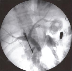
Figure 1 Intraoperative cholangiogram via the cystic duct demonstrating proximal biliary dilation and two filling defects in the CBD (Arrows).
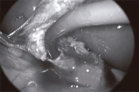
Figure 2 Laparoscopic view of a choledochoscope (CS) entering the CBD via the cystic duct.
The gallbladder (GB) is retracted to the left of the image.
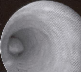
Figure 3 CBD stone as seen through the choledochoscope.
- Citation: Freitas ML, Bell RL, Duffy AJ. Choledocholithiasis: Evolving standards for diagnosis and management. World J Gastroenterol 2006; 12(20): 3162-3167
- URL: https://www.wjgnet.com/1007-9327/full/v12/i20/3162.htm
- DOI: https://dx.doi.org/10.3748/wjg.v12.i20.3162


