Copyright
©2006 Baishideng Publishing Group Co.
World J Gastroenterol. Mar 14, 2006; 12(10): 1634-1636
Published online Mar 14, 2006. doi: 10.3748/wjg.v12.i10.1634
Published online Mar 14, 2006. doi: 10.3748/wjg.v12.i10.1634
Figure 1 Colonoscopic examination of early minimal lesion (A) and advanced lesion (B) showing slightly elevated bright red round patches and congestive hyperemic polypoid lesion contrasted sharply with the surrounding mucosa (B: indigo carmine dye spraying view).
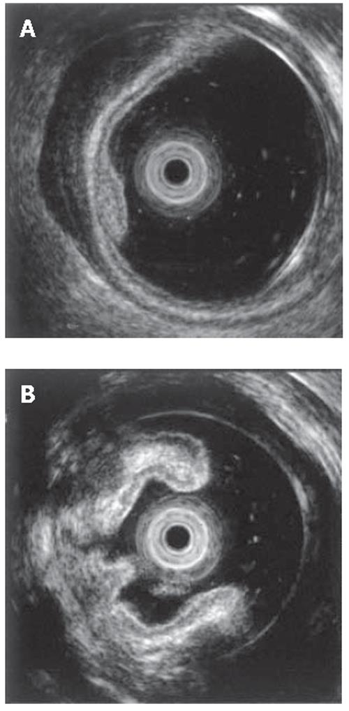
Figure 2 Endoscopic ultrasonographic examination of early minimal lesion (A) and advanced lesion (B) showing thickening of the first and second layers (mucosa) and irregular echoic lesion in the third layer (submucosa) with hypertrophy of the fourth layer (muscularis propria), and irregular low echoic lesion throughout colonic wall, respectively.
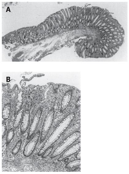
Figure 3 Hisological examination of prolapsing mucosal polyp showing mucosal hyperplasia (A) and chronic inflammatory cell infiltration, crypt elongation, congested vessels and fibromuscular obliterations observed in the lamina propria (B) (H&E, a: x40, b: x200).
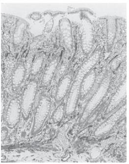
Figure 4 α-Smooth muscle actin (α-SMA) staining of prolapsing mucosal polyp showing smooth muscle fibers radiating from the thickened muscularis mucosa into the lamina propria (x200).
- Citation: Kato S, Hashiguchi K, Yamamoto R, Seo M, Matsuura T, Itoh K, Iwashita A, Miura S. Jumbo biopsy is useful for the diagnosis of colonic prolapsing mucosal polyps with diverticulosis. World J Gastroenterol 2006; 12(10): 1634-1636
- URL: https://www.wjgnet.com/1007-9327/full/v12/i10/1634.htm
- DOI: https://dx.doi.org/10.3748/wjg.v12.i10.1634


