©2006 Baishideng Publishing Group Co.
World J Gastroenterol. Jan 7, 2006; 12(1): 54-59
Published online Jan 7, 2006. doi: 10.3748/wjg.v12.i1.54
Published online Jan 7, 2006. doi: 10.3748/wjg.v12.i1.54
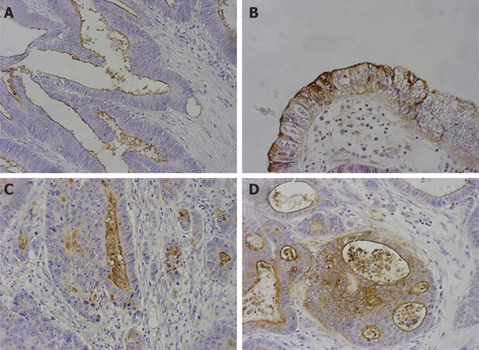
Figure 1 Immunohistochemical staining of colorectal adenocarcinoma tissues using KL - 6 antibody (×200).
A: Positive staining in the apical membrane; B: positive staining in the circumferential membrane; C: positive staining in the apical membrane and cytoplasm; D: positive staining in the circumferential membrane and cytoplasm.
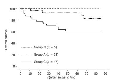
Figure 2 Kaplan - Meier curves for overall survival rates of patients with colorectal adenocarcinoma.
Patients with KL - 6 expression in the circumferential membrane and/or cytoplasm (solid line, group C, n = 46), in the apical membrane (dashed line, group A, n = 29) and without KL - 6 staining (dotted line, group N, n = 5) were followed up for more than 70 mo. Two of 82 patients were excluded from the data analysis as described in Materials and Methods.
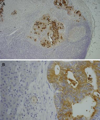
Figure 3 Immunohistochemical staining of metastatic lymph node and liver tissues using KL-6 antibody (×200).
A:Positive KL-6 expression in a metastatic lymph node; B: positive KL-6 expression in metastatic liver tissues.
- Citation: Guo Q, Tang W, Inagaki Y, Midorikawa Y, Kokudo N, Sugawara Y, Nakata M, Konishi T, Nagawa H, Makuuchi M. Clinical significance of subcellular localization of KL-6 mucin in primary colorectal adenocarcinoma and metastatic tissues. World J Gastroenterol 2006; 12(1): 54-59
- URL: https://www.wjgnet.com/1007-9327/full/v12/i1/54.htm
- DOI: https://dx.doi.org/10.3748/wjg.v12.i1.54


