©2006 Baishideng Publishing Group Co.
World J Gastroenterol. Jan 7, 2006; 12(1): 43-47
Published online Jan 7, 2006. doi: 10.3748/wjg.v12.i1.43
Published online Jan 7, 2006. doi: 10.3748/wjg.v12.i1.43
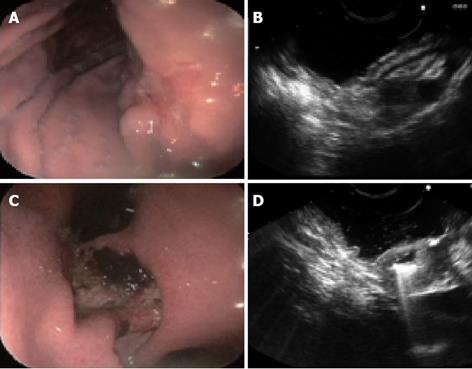
Figure 1 Early and advanced gastric cancer cases.
A: Endoscopic view of superficial depressed type of early gastric cancer; B: EUS image shows cancer invasion of 1st and 2nd (mucosal) layers of gastric wall, while 3rd (submucosal) layer is clear (T1 category). Histopathological findings of the surgically resected specimen corresponded with the EUS findings; C: Endoscopic view of advanced Borrmann II type of gastric cancer; D: EUS images show disruption of 1-4 layers of the gastric wall with hypoechoic cancer tissue, but 5th (serosal) layer is not involved (T2 category).
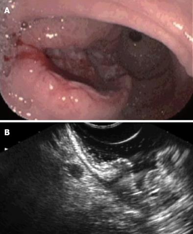
Figure 2 A case of advanced gastric cancer.
A: Endoscopic view of Borrmann III type of gastric cancer; B: EUS image demonstrates T3 cancer with malignant lymph node. Note the hypoechoic structure and sharp margin of the lymph node (1.0 cm×0.6 cm).
- Citation: Tsendsuren T, Jun SM, Mian XH. Usefulness of endoscopic ultrasonography in preoperative TNM staging of gastric cancer. World J Gastroenterol 2006; 12(1): 43-47
- URL: https://www.wjgnet.com/1007-9327/full/v12/i1/43.htm
- DOI: https://dx.doi.org/10.3748/wjg.v12.i1.43


