Copyright
©2005 Baishideng Publishing Group Inc.
World J Gastroenterol. Feb 28, 2005; 11(8): 1149-1154
Published online Feb 28, 2005. doi: 10.3748/wjg.v11.i8.1149
Published online Feb 28, 2005. doi: 10.3748/wjg.v11.i8.1149
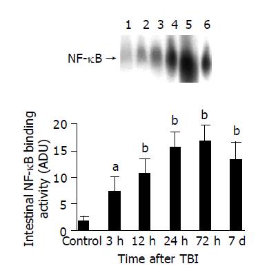
Figure 1 Jejunal NF-κB binding activity following TBI.
As compared with control group, NF-κB binding activity in the jejunal tissue following TBI significantly increased at 3 h after TBI, being highest at 72 h postinjury, and remained elevated by 7 d postinjury. Upper: EMSA autoradiogram. Lane 1, control; lane 2, 3 h postinjury; lane 3, 12 h postinjury; lane 4, 24 h postinjury; lane 5, 72 h postinjury; lane 6, 7 d postinjury. Lower: bar graph showing the mean band intensity of EMSA autoradiograph in the jejunal tissue of control, 3, 12, 24, 72 h and 7 d postinjury. aP<0.05, bP<0.01. mean±SD of six animals in each group.
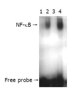
Figure 2 Results of competitive electrophoretic mobility shift assay for NF-κB activity.
Lane 1, negative control, no Hela nuclear extract; lane 2, positive control, Hela nuclear extract; lane 3, Hela nuclear extract plus 100-fold molar excess of unlabeled NF-κB consensus oligo (specific competitor); lane 4, Hela nuclear extract plus 100-fold molar excess of unlabeled AP2 consensus oligo (nonspecific competitor). This autoradiograph is representative of 2 independent experiments.
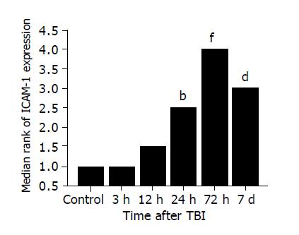
Figure 3 Expression intensity of ICAM-1 in jejunum, manifested as median rank.
There was no significant difference in the ICAM-1 positive cells among the rats of control group, TBI 3 h and 12 h. As compared to controls, ICAM-1 immunoreactivity was significantly up-regulated on the endothelia of microvessels in villous interstitium and lamina propria by 24 h following TBI and maximally expressed at 72 h postinjury. The endothelial ICAM-1 expression in jejunal interstitium still remained high at 7 d postinjury. bP = 0.01 vs control, dP = 0.001 vs control, fP<0.001 vs control.
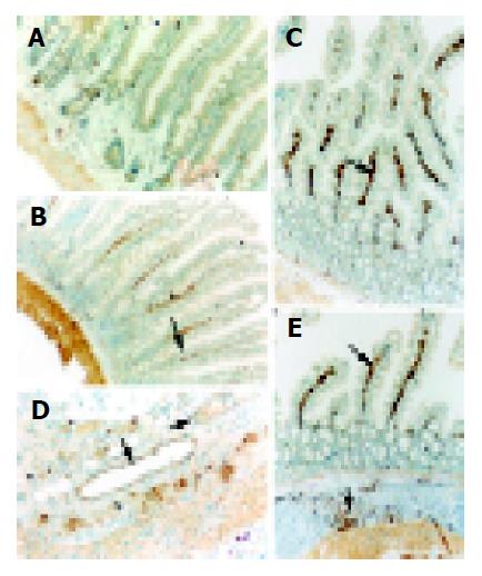
Figure 4 ICAM-1 immunohistochemical staining of jejunal tissue, indicated by the arrows.
A: almost undetectable ICAM-1 expression in the villi from control rats; B: moderate ICAM-1 immunoreactivity in the villous interstitium at 24 h postinjury, with the villi well defined; C: strong ICAM-1 immunoreactivity in the villous interstitium at 72 h postinjury, with the villi disarranged and the ICAM-1 positive endothelial cells shown as nigger-brown granules; D: marked ICAM-1 immunohistochemical staining of microvessels in the intestinal lamina propria at 72 h postinjury; E: high ICAM-1 immunoreactivity in the villous interstitium at 7 d postinjury, with the villi atrophic and sparse.
- Citation: Hang CH, Shi JX, Li JS, Li WQ, Yin HX. Up-regulation of intestinal nuclear factor kappa B and intercellular adhesion molecule-1 following traumatic brain injury in rats. World J Gastroenterol 2005; 11(8): 1149-1154
- URL: https://www.wjgnet.com/1007-9327/full/v11/i8/1149.htm
- DOI: https://dx.doi.org/10.3748/wjg.v11.i8.1149


