Copyright
©The Author(s) 2005.
World J Gastroenterol. Jul 21, 2005; 11(27): 4281-4284
Published online Jul 21, 2005. doi: 10.3748/wjg.v11.i27.4281
Published online Jul 21, 2005. doi: 10.3748/wjg.v11.i27.4281
Figure 1 A: Liver CT scan showed a huge tumor in the right lobe of the liver with enhancement from the periphery in the arterial phase; B: The hepatic arteriogram demonstrated numerous feeding vessels surrounding and entering into the tumor.
The patient had variation of the right hepatic artery from the superior mesenteric artery.
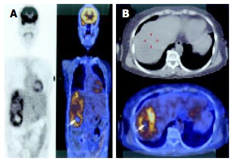
Figure 2 A: 18F-FDG PET showed high uptake at the tumor without any distant lesions.
The silent area in the center of the tumor suggests central necrosis (arrow); B: The highest uptake area measured 4.8 SUV (red line mark).
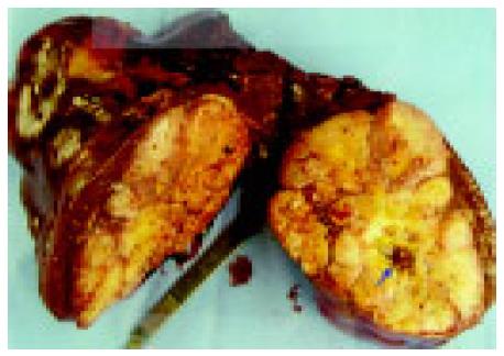
Figure 3 Resected specimen showed a huge single tumor with sarcomatous consistency.
The margin was irregular because of the constrictive envelopment of feeding vessels. The central necrosis is indicated (arrow).
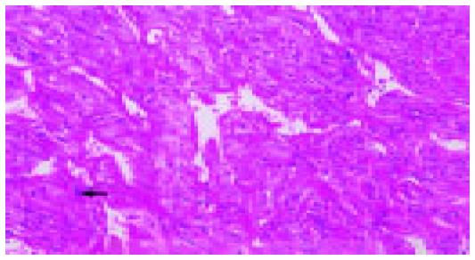
Figure 4 Microscopic examination demonstrated numerous atypical vessels (stag-horn appearance) in the high population of spindle-shaped tumor cells.
A mitotic figure is seen (arrow), H&E, ×200.
- Citation: Kim BW, Wang HJ, Jeong IH, Ahn SI, Kim MW. Metastatic liver cancer: A rare case. World J Gastroenterol 2005; 11(27): 4281-4284
- URL: https://www.wjgnet.com/1007-9327/full/v11/i27/4281.htm
- DOI: https://dx.doi.org/10.3748/wjg.v11.i27.4281


